What is percutaneous kidney biopsy?
A kidney biopsy, also called renal biopsy, is a procedure to remove a small piece of kidney tissue so that it can be examined under a microscope for signs of damage or disease. Your doctor may recommend a kidney biopsy if blood tests, urine tests or imaging techniques don’t provide enough information to diagnose a kidney problem. A kidney biopsy may also be used to determine the severity of kidney disease or to find out if treatment for kidney disease is working. You may need a kidney biopsy if you’ve had a kidney transplant that’s not working properly.
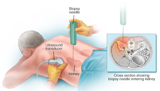
A kidney biopsy is done by inserting a long thin needle through the back (flank) into the kidney. This is called a percutaneous kidney biopsy. Ultrasound or Computed Tomography (CT) is often used to help guide the interventional radiologist’s instruments to the site of the kidney.
What are some common uses of the procedure?
Some common reasons for kidney biopsy include:
- Blood in the urine (haematuria);
- Protein in the urine (proteinuria) that’s excessive, rising or accompanied by other signs of kidney disease;
- Problems with kidney function, leading to excessive waste products in the blood.
Not everyone with these problems needs a kidney biopsy. The decision is based on your signs and symptoms, test results and overall health.
How should I prepare?
You may be instructed not eat or drink for eight hours before your biopsy. However, you may take your routine medications with sips of water.
Prior to a needle biopsy, you should report to your doctor all medications that you are taking, and if you have any allergies, especially to anaesthesia. Your doctor will advise you to stop taking aspirin or a blood thinner three days before your procedure.
Also, inform your doctor about recent illnesses or other medical conditions.
In your ward, before being taken to the Imaging Department, you may be asked to remove some or all of your clothes, to wear a hospital gown and an intravenous line will be placed.
In case of CT-guided biopsy, you may also be asked to remove jewellery and any metal objects or clothing that might interfere with the X-ray transmission. Women should always inform their doctor if there is any possibility that they are pregnant. Some procedures using imageguidance are typically not performed during pregnancy because radiation can be harmful to the foetus. If an X-ray is necessary, precautions will be taken to minimise radiation exposure to the baby.
You will have blood tests done before the kidney biopsy to see whether you have any bleeding problems or blood clotting disorders.
It is recommended to have a relative or friend accompany you and drive you home afterward.
How is the procedure performed?
Imaging-guided, minimally invasive procedures such needle kidney biopsy are most often performed by a specially trained interventional radiologist.
Your kidneys lie just under the ribcage, towards the side and back of your upper abdomen. So, you will usually be asked to lie on your front on a couch or bed. The skin over a kidney is cleaned with antiseptic and a clean, sterile drape will be applied. Local anaesthetic is then injected into a small area of skin and tissues just over the kidney to be biopsied. The doctor then makes a small incision where the needle will go in. An ultrasound or CT device is used to guide the needle into the correct place in your kidney.
You’ll be asked to stop breathing as the doctor inserts the biopsy needle into the kidney. When the needle pushes through your skin to the kidney, you may feel some pressure and hear a sharp clicking noise from the sampling instrument. You’ll remain still and hold your breath for the time it takes to collect the tissue, 20 to 30 seconds. Your doctor may need to insert the needle two or three times (often through the same incision) to get enough tissue.
Once the biopsy is complete, pressure will be applied to stop any bleeding and the opening in the skin is covered with a dressing. No sutures are needed. This procedure is usually completed within one hour.
You will return to your ward and at least six hours later, abdominal ultrasound will be performed to monitor for complications.
What will I experience during and after the procedure?
When you receive the local anaesthetic to numb the skin, you will feel a slight pin prick from the needle. This stings a little at first, but then, the area will become numb within a short time.
You will be asked to remain still during the procedure. You also will be asked to hold your breath multiple times during the biopsy. It is important that you try to maintain the same breath-hold each time to insure proper needle placement.
Generally your bandage may be removed one day following the procedure and you may bathe or shower as normal.
You should not exert yourself physically (such as heavy lifting, extensive stair climbing, sports, etc.) for one week following your biopsy. You will return to your normal activities soon. You may eat your normal diet. Do not take aspirin for a week after the biopsy.
You may experience some soreness at the biopsy site as the local anaesthesia fades, but this should improve. Many people will have bright red blood in urine for the first 24 hours after the biopsy; this is expected.
In some cases, bleeding may occur, originating from the kidney itself. Sometimes, surgery is needed to control bleeding. You will be charged for all extra treatments.
Who interprets the results and how do I get them?
A pathologist examines the removed specimen and makes a final diagnosis so that treatment planning can begin. Depending on the facility, the radiologist or your referring doctor will disclose the results to you.
What are the benefits and risks?
Benefits
- Diagnose a kidney problem that can’t otherwise be identified.
- Help develop treatment plans based on the kidney’s condition.
- Recovery time is brief and patients can soon resume their usual activities.
Risks
- Any procedure where the skin is penetrated carries a risk of infection. The chance of infection requiring antibiotic treatment appears to be less than one in 1,000.
- In general, percutaneous kidney biopsy is a safe procedure.
- The most common complication of kidney biopsy is blood in the urine (haematuria). The bleeding usually stops within 24 hours. Bleeding that’s serious enough to require a blood transfusion affects a very small percentage of people who have a kidney biopsy. Rarely, surgery is needed to control bleeding.
- If the biopsy needle accidentally damages the walls of a nearby artery and vein, a fistula, or abnormal connection, can form between the two blood vessels. This type of fistula usually causes no symptoms and closes on its own.
- Rarely, a collection of blood (haematoma) around the kidney becomes infected. This complication is treated with antibiotics and surgical drainage.
IMAGING DEPARTMENT
FV Hospital, Ground Floor, F building
Tel: (028) 54 11 34 00