WHAT IS ULTRASOUND-GUIDED BREAST BIOPSY?
Lumps or abnormalities in the breast are often detected by physical examination, ultrasound, mammography, or other imaging studies. However, it is not always possible to tell from these imaging tests whether a growth is benign or cancerous.
A breast biopsy is performed to remove some cells—either surgically or through a less invasive procedure involving a hollow needle—from a suspicious area in the breast and examine them under a microscope to determine a diagnosis.
Image-guided biopsy is performed when the abnormal area in the breast is too small to be felt, making it difficult to locate the lesion by hand (called palpation).
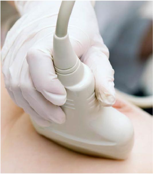
In ultrasound-guided breast biopsy, ultrasound imaging is used to help guide the radiologist’s instruments to the site of the abnormal growth.
WHAT ARE SOME COMMON USES OF THE PROCEDURE?
An ultrasound-guided breast biopsy can be performed when a breast ultrasound shows an abnormality such as:
- A suspicious solid mass
- A distortion in the structure of the breast tissue
- An area of abnormal tissue change
Ultrasound guidance is used in three biopsy procedures:
- Fine needle aspiration (FNA), which uses a very small needle to extract cells from the abnormal area.
- Core needle, which uses a large hollow needle to remove one sample of breast tissue per insertion.
- Wire localization, in which a guide wire is placed into the suspicious area to help the surgeon locate the lesion for surgical biopsy.
HOW SHOULD I PREPARE?
You will be asked to fill out a Questionnaire and a Consent form before the exam.
You will need to remove all clothing and jewellery in the area to be examined.
Prior to a needle biopsy, you should report to your doctor all medications that you are taking, and if you have any allergies, especially to local anaesthesia. Your doctor will advise you to stop taking blood thinning medication (anticoagulant medication) three days before your procedure.
You should also, inform your doctor about recent illnesses or other medical conditions.
It is recommended to have a relative or friend accompany you and drive you home afterward.
HOW IS THE PROCEDURE PERFORMED?
Breast biopsies are usually done on an outpatient basis.
You will be positioned lying face up on the examination table or turned slightly to the side.
The lesion is immediately visible on a nearby screen as the ultrasound probe passes over the concerned area. After locating the site of concern, the skin is cleansed. If a FNA is indicated, no anaesthesia is administered. A local anaesthetic will be injected in the case of core biopsy.
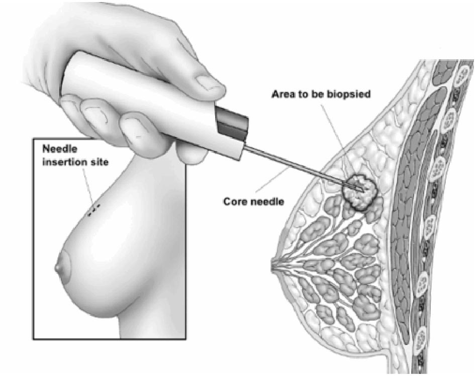
A very small nick may be made in the skin at the site where the biopsy needle is to be inserted.
The radiologist, constantly monitoring the lesion site with the ultrasound probe, will insert the needle and advance it directly into the mass. As tissue samples are taken, you may hear clicks from the sampling instrument. After this sampling, the needle will be removed. Tissue samples will then be removed.
This process is repeated three times or more.
Once the biopsy is complete, pressure will be applied to stop any bleeding and the opening in the skin is covered with a dressing.
WHAT WILL I EXPERIENCE DURING AND AFTER THE PROCEDURE?
You will be awake during your biopsy and should have little or no discomfort. Most women report little or no pain and no scarring on the breast.
When you receive the local anaesthetic to numb the skin, you will feel a slight pin prick from the needle. You may feel some pressure when the biopsy needle is inserted.
You must remain still while the biopsy is performed.
If you experience swelling and bruising following your biopsy, you may be instructed to take an over-the-counter pain reliever. Temporary bruising is normal.
You should contact your doctor if you experience excessive swelling, bleeding, drainage, redness or heat in the breast.
HOW LONG DOES AN ULTRASOUND-GUIDED BREAST BIOPSY TAKE?
This procedure is usually completed within an hour.
How about the results?
A pathologist in the laboratory will examine the removed specimen and will make a diagnosis. Results are usually given within a few working days.
What are the benefits vs. risks?
Benefits
- The procedure is less invasive than surgical biopsy, leaves little or no scarring and can be performed in less than an hour.
- Ultrasound-guided breast biopsy reliably provides tissue samples that can show whether a breast lump is benign or malignant.
- The ultrasound method avoids the need for ionizing radiation exposure.
- With ultrasound it is possible to follow the motion of the biopsy needle as it moves through the breast tissue.
- Ultrasound-guided breast biopsy is able to evaluate lumps located deep or near the chest wall, which are hard to reach with stereotactic biopsy.
- Ultrasound-guided biopsy is less expensive but faster than stereotactic biopsy.
- Recovery time is brief and patients can soon resume their usual activities.
Risks
- Any procedure where the skin is penetrated carries a risk of infection. The chance of infection requiring antibiotic treatment appears to be less than one in 1,000.
- Doing a biopsy of tissue located deep within the breast carries a slight risk that the needle will pass through the chest wall, allowing air around the lung that could collapse a lung.
- This is a rare occurrence.
WHAT ARE THE LIMITATIONS OF ULTRASOUND-GUIDED BREAST BIOPSY?
Breast biopsy procedures will occasionally underestimate the extent of disease present. If the diagnosis remains uncertain after a technically successful procedure, surgical biopsy will usually be necessary.
The ultrasound-guided biopsy method cannot be used unless the lesion can be seen on an ultrasound exam. Clustered calcifications are not shown as clearly with ultrasound as with x-rays and therefore, stereotactic biopsy is indicated.
Very small lesions may be difficult to target accurately by ultrasound-guided core biopsy.
HOW DO I SCHEDULE AN ULTRASOUND-GUIDED BREAST BIOPSY?
For further information and for taking appointment, kindly contact our imaging department at (08) 54 11 34 00