Technological breakthroughs in imaging are helping physicians, in all fields of medicine, to diagnose and treat diseases faster and better than ever before. Today, nuclear medicine is opening the door into the human body, and, in most cases, eliminating the need for exploratory surgery.
Before the advent of nuclear medicine, patients often had to undergo exploratory surgery so that doctors could check for tumors and evaluate the progress of chemotherapy treatment. Such surgeries often involved significant risk, recovery time and cost.
At FV Hospital, nuclear scanning is widely used to diagnose heart disease, thyroid conditions and kidney tumors, and to assess patients with cancers, because it provides physicians with fast and detailed images for more accurate diagnosis and treatment.

Nuclear scanning
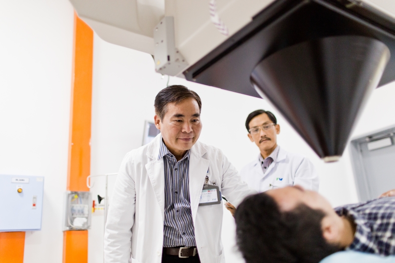
Nuclear scanning sounds scary, but it is really quite simple and safe. The objective is to illuminate tissue so that the imaging device can see what is happening inside the body. The nuclear scan starts with a small amount of radioactive agent injected into the vein. Once this radioactive material enters the bloodstream, it settles into body tissue making it visible to the scanning device. The scan itself can take from a few minutes to a few hours to complete, and no anaesthetic is required. The amount of radiation received from a nuclear scan is extremely small, about the same as from a standard x-ray, and afterwards it is excreted naturally from the body.
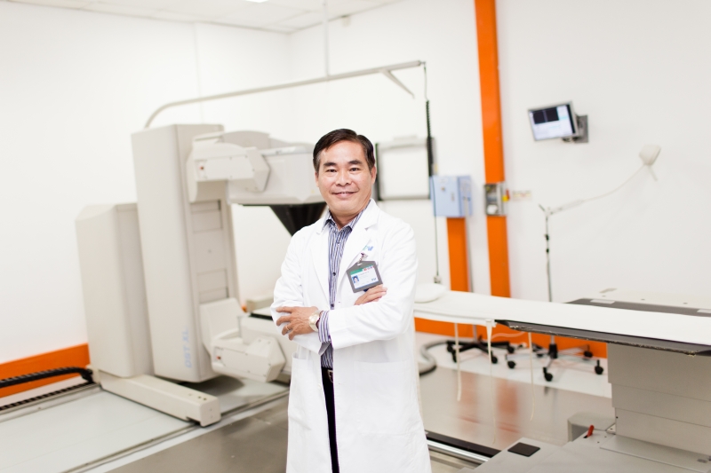
The Gamma Camera

The gamma camera is used to produce images of organs or tissue and seeks out radiation emitted by the radioactive material. It is highly sensitive to radiation (but it does not emit radiation) and essentially shows where the radioactive material is settling inside the patient’s body. This gives the doctor valuable information to help determine the right course of treatment or whether a particular treatment is working. The camera is capable of taking a full body scan or can concentrate on a particular area or organ, depending on what the doctor needs to see. The camera can also produce three-dimensional images that help the doctor study the problem from every possible angle.
“At FV Hospital, using the newest generation of gamma cameras, we are able to work with very clear and precise images”, said Dr. Nguyen Van Te, Director of the Nuclear Medicine Department, “Enabling our doctors to make faster, more precise diagnoses, leading to better treatment. In the end, that benefits everyone.”
No results found...


