FV Hospital has acquired the world’s most advanced hybrid SPECT/CT system to offer cutting edge diagnostic and therapeutic capabilities in the field of Nuclear Medicine. This is a 2-in-1 device which combines single-photon emission computed tomography (SPECT) using gamma rays and computed tomography (CT) cameras. This system allows physicians to proactively capture 2D images or 2D and 3D scans for overall imaging purposes, along with corrected radiographic degradation and detailed description of diseases across the whole body or in a specific area.
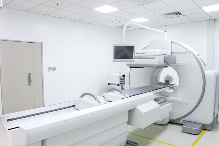
The SPECT/CT system is non-invasive, painless and non-allergenic and has side effects. FV Hospital is one of the few hospitals in the country to own a SPECT/CT system.
HOW A SPECT/CT DEVICE WORKS
Before a SPECT scan, patients are usually injected with the artificial radioactive isotope 99m Tc (which as a short half-life of six hours) attached to a drug specific for each organ that is to be examined. Depending on the case, radioactive substance can be introduced into the body via the mouth, it can be inhaled or delivered as subcutaneous injection. In addition, patients may be given 131 Iodine, another artificial radioactive isotope with a short half-life of eight days.
These compounds emit Gamma rays. Two detectors receive these rays simultaneously from the patient’s body and specialised software produces images.
Patients will be instructed by the operator to lie on the table within the scan range of the SPECT/CT scanner to achieve the images required by their doctors. Scan times typically range from 15 minutes to 45 minutes or longer, depending on the complexity of the location of the lesion (there are times when an area needs re-scanning, which results in a longer period spent on the table). After the scan is complete, the patient may be asked to remain in the hospital for a short period of time or may be discharged immediately.
After the image is processed, the results highlight lesions in the body in the form of anatomical and functional images. In complex cases, Nuclear Medicine doctors will discuss the results with the doctor who has requested an SPECT/CT scan. Images and reports will then be handed to the patient or sent directly to their doctor.
Directions to be strictly followed:
- Before the scan, the patient must be sure that she is not pregnant. If unsure, the patient should immediately inform employees or doctors (blood examination may be necessary to confirm the pregnancy).
- Women who are breastfeeding need to stop breastfeeding within 12 hours to 24 hours prior to the scan.
- There are no contraindications for children.
- Usually, it is highly recommended for patients to drink plenty of water before being injected with a radioactive drug.
- Patients may need to make an appointment for another consultation with their doctor.
Doctors will prescribe a SPECT/CT scan in the following cases:
Cardiovascular disease: A review of myocardial ischemia and left ventricular function in patients with previous coronary artery disease, or patients experiencing chest pain to detect coronary artery disease.
Tumours:
– A complete bone scan will be ordered to find metastatic lesions and evaluate the progression of metastatic cancer during the course of the disease.
– Full body radiography with 131 Iodine:
- Postoperative thyroidectomy
- Before and after 131 iodine treatment
- Monitor treatment response in differentiated thyroid gland cancer.
– Lymphoscintigraphy for detection of Sentinel Lymph node (in early breast cancer, melanoma and some other cancers in the head, face and neck, etc.) help assess the stage and treatment plan.
Endocrinology:
– To facilitate diagnosis of thyroid gland diseases (including hyperthyroidism, hepatomegaly, thyroid tumours, thyroid cancer, etc.).
– To detect adrenal tumours, neuroblastoma, parathyroid gland (parathyroid), etc.
Urology:
– In kidney radiation (static):
- To help assess damage to the renal cortex and the remaining function of each kidney, so that doctors can decide whether to treat or surgically remove all or part of the kidney.
- To find the location of a misplaced kidney.
– In kidney radiation (motion):
- To evaluate individual functions of each kidney while diagnosing and differentiating kidney stagnancy due to obstruction or non-obstruction.
- To diagnose hypertension due to renal artery stenosis.
- To assess renal function before donation and after renal transplant.
Testicular imaging helps to distinguish inflamed orchitis and testicular torsion.
Orthopaedic:
– To diagnose an infection or a loose joint.
– To support diagnosis of the cause of bone pain, suspected osteoarthritis, or bone marrow problems.
ICU/Emergency:
– To perform pulmonary radiography and perfusion: the diagnosis will determine or exclude pulmonary embolism.
Other cases:
– To radiographically identify Meckel’s diverticulum, salivary glaucoma.
The new SPECT/CT system is an additional diagnostic and pathological support tool for FV Hospital doctors, improving their ability to provide a more accurate assessment of the function and surgical site of the lesion than previous generation technologies.