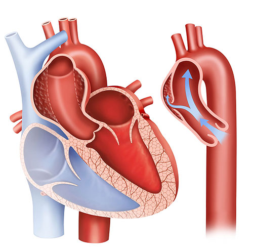
In mid-April, the A&E Department of FV Hospital received a French patient, L.M (52 years old) who was hospitalized with severe back pain. The pain occurred suddenly and then gradually spread to the abdomen area. Through a combination of examination and history of hypertension of the patient, doctors suspected that the symptoms were related to thoracic aortic dissection. The CT scan results showed that inside the thoracic aorta appeared an ablation from the lower arterial artery on the left, which had spread to the pelvic arteries on either side. The thoracic aorta was split in half, creating a false vein, which causes compression of the vascular lumen, and in turn reduces the amount of blood flowing to the arteries that feed the organs (intestine, liver, kidney, among others). This phenomenon in medical term is called type B thoracic aortic dissection, usually occurring to older people with a history of atherosclerosis and high blood pressure…
“Mr L.M was admitted to the hospital right after the detection of abnormal signs, when the dissection situation did not cause danger to the organs and could control the blood pressure by medicine”, shared Doctor Luong Ngoc Trung, the Thoracic & Vascular Surgeon, who directly conducted treatment for the patient. The complication had not occurred yet, so it could be postponed with medical treatment and be followed-up in the intensive care unit. However, due to the patient’s thinking that “the disease was not serious”, the patient was determined to return to France to receive treatment there. “At that time, Mr L.M’s health was not good enough to endure a long flight from Vietnam to France. Not to mention, when the aircraft took off and landed, the air pressure changed, raising blood pressure, causing an artery rupture, or causing the disease to turn into complication of upstream Aortic dissection, which, in some cases, might lead to death”, Dr Trung explained. After that, FV Hospital’s CEO, Dr Jean-Marcel Guillon, personally met and persuaded the patient to be assured of the quality of FV’s standards of treatment. Despite this, Mr L.M’s hesitation still existed. On the third day, the patient’s condition began to worsen when the pain in the chest and abdomen became less responsive to painkillers. The results of the second CT scan showed that the false lumen had enlarged more than before, the medical treatment with medicine was no longer effective.

Dr Luong Ngoc Trung
If not timely surgery was administered; it would lead to the risk of complications.
Considering the level of seriousness of the situation, the patient agreed to the treatment procedure by placing a stent-graft.
Accordingly, the doctors conducted intravascular intervention by opening a small incision in the groin, classifying the true and false veins to put the stent into the heart. Along with the support from the state-of-the-art Cathlab room, invested by FV in accordance with international standards, Dr Luong Ngoc Trung cleverly positioned the 3 stent-graft in the right place within the damaged artery. The surgery lasted about 2 and a half hours and was a huge success. The results of postoperative imaging showed that the shape and flow of the damaged aortic artery was well restored, the blood vessels supplying the organ were back to normal. The patient’s health turned stable a few days later and no instances of further complications arose.
FV Hospital is equipped with advanced imaging diagnostic systems, especially the latest 256-MSCT equipment, which helps doctors quickly and accurately diagnose cardiovascular diseases. Previously, FV successfully treated many cases of large-sized aortic aneurysms in elderly patients with many complex conditions.
Thoracic & Vascular Surgery Department – 3rd floor, V building, FV Hospital
To make an appointment, please contact: 028 5411 3333