In cases of coronary restenosis, the biggest challenge for physicians lies in accurately assessing the intricate damage within the coronary arteries to devise precise treatment plans, as treatment outcomes can be greatly affected. However, with the intervention of Optical Coherence Tomography (OCT) imaging technology, every nook and cranny within the arteries is vividly depicted, greatly enhancing the treatment of coronary arteries, particularly in cases of restenosis, resulting in highly successful outcomes.
Therefore, OCT is deemed an invaluable aid for cardiologists, offering the potential to save the lives of numerous patients experiencing acute coronary artery restenosis.
Placing a stent for patients experiencing coronary artery restenosis is facilitated by “guidance” provided by OCT (Optical Coherence Tomography) technology.
Mr N., a German patient, was brought to FV Hospital’s emergency department by his family in November 2023, presenting symptoms of chest pain and difficulty in breathing. Following diagnostic tests, the Cardiology Department concluded that Mr N. was suffering from restenosis in the right coronary artery. Five years ago, he had undergone coronary artery stent intervention in Germany. However, new atherosclerotic plaques had formed, narrowing the artery and directly affecting the heart’s function and performance. The patient might encounter severe complications such as arrhythmias, heart failure, myocardial infarction, among others, if not promptly treated.
To thoroughly assess Mr N.’s coronary artery damage before devising a treatment plan, the medical team decided to utilise the newly implemented OCT imaging system for coronary arteries at FV Hospital.
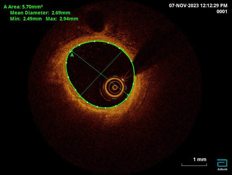
The OCT imaging clearly depicts the lesions within the coronary arteries
OCT for coronary arteries is a cutting-edge imaging technique. Physicians insert an incredibly small imaging device into the coronary arteries, emitting short-wavelength infrared light to capture images within the arteries. These images are highly detailed, allowing for a clear view of the structures within the arteries, such as the layers of blood vessels, ulcers, ruptures, blood clots, and more. As a result, doctors can precisely identify the location for stent placement, select an appropriately sized stent, and reduce the risk of restenosis.

The OCT (Optical Coherence Tomography) technique is employed by the doctors at FV Hospital to treat patients with coronary artery restenosis.
From the precise, detailed imaging results, the team led by Ho Minh Tuan, MD, PhD, proposed a treatment plan for coronary artery restenosis in patient N. This plan involved removing atherosclerotic plaques using a cutting balloon and ablating calcified plaques with a diamond-tipped burr, followed by enlarging the narrowed artery and implanting a new stent. Prior to concluding the procedure, the physicians rechecked the newly placed stent using OCT to ensure its accurate positioning and optimal expansion. Subsequently, the patient recovered well and was discharged from the hospital.
OCT technology supports an effective treatment for patients with coronary artery restenosis.
Coronary artery disease results from the accumulation of atherosclerotic plaques on the arterial walls, causing the arteries to narrow over time. This narrowing progresses from mild to severe and can occasionally lead to complete blockages. The condition can cause silent myocardial ischemia, angina (unstable angina, myocardial infarction), and sudden cardiac death. According to statistics from the National Heart Institute, the incidence of coronary artery disease is steadily increasing each year, accounting for 11-36 per cent of mortality rates.
Conventional stent placement for coronary artery treatment often utilises Digital Subtraction Angiography (DSA) imaging for blood vessel and coronary angiography. However, this method has limitations in displaying hard-to-see lesions, requiring multiple X-ray exposures, potentially affecting the patient’s health. Another technique, IVUS (Intravascular Ultrasound), uses high-penetration ultrasound waves to visualise atherosclerotic plaques within the arteries. Nevertheless, IVUS images have limited resolution and are mainly applied in larger blood vessels. With these two techniques, physicians may struggle to accurately assess complex lesions, which can impact treatment outcomes.
“Estimates suggest that around 25-30 per cent of cases diagnosed with coronary artery stenosis and stent placement interventions might not be optimised without OCT technology. The introduction of OCT for coronary arteries has addressed these issues and created opportunities for better treatment for patients with coronary artery restenosis and other cardiovascular conditions,” states Ho Minh Tuan, MD, PhD, Head of the Cardiology Department at FV Hospital.
Clinical studies have shown that OCT technology significantly aids physicians in effectively treating cases of coronary artery restenosis. “Firstly, OCT helps identify the reasons behind a patient’s restenosis and then assesses the optimisation of stent placement. Secondly, in cases of branched coronary lesions, OCT clearly depicts these lesions, enabling physicians to devise accurate treatment plans. Branch lesions are considered complex injuries, requiring OCT for precise analysis in treatment,” noted the Head of the Cardiology Department at FV Hospital.

Ho Minh Tuan, MD, PhD says that in terms of intravascular imaging for coronary arteries, OCT is the most advanced technique currently available.
OCT for coronary arteries is highly regarded by medical professionals in cardiac interventions, as it enhances treatment efficacy and safety for patients. However, to operate this technique, hospitals require a specialised team of trained physicians, technicians, and modern equipped catheterization labs (Cathlab).
For more information about Optical Coherence Tomography (OCT) imaging technology, please contact: (028) 35 11 33 33.