Table of contents
WHAT IS FNA?
Fine Needle Aspiration (FNA) is a diagnostic procedure used to investigate superficial (just under the skin) lumps or masses. Your radiologist, under ultrasound guidance, will attempt to retrieve enough tissue for microscopic analysis and thus make a diagnosis of a number of problems, such as inflammation, infection, benign tumour or even cancer. This procedure often, but not always, prevents the patient from having an open, surgical biopsy.
WHY IS FNA IMPORTANT?
A lump (or ‘mass’), a nodule (lumps in the thyroid gland are called ‘nodules’) sometimes indicates a serious problem, such as cancer; but a mass or a nodule can be benign (non-cancerous) or related to an infection. The presence of a mass or a nodule may require FNA for diagnosis.
Other factors are taken in consideration by your doctor for the diagnosis: your age, sex, and habits, such as smoking and drinking; symptoms of ear pain, increased difficulty swallowing, weight loss; a history of familial thyroid disorder or of previous skin cancer (squamous cell carcinoma).
Note that when found early, most cancers in the head and neck can be cured. So if you have a lump in your head and neck area, see your Oto-Rhino-Laryngologist (ORL, also called ‘ENT’ for Ear, Nose and Throat) right away.
WHAT ARE THE AREAS THAT CAN BE INVESTIGATED BY FNA?
FNA is generally used for diagnosis of neck lymph nodes and cysts. FNA is the most commonly performed test to determine whether a thyroid nodule is benign or suspicious for malignancy. The parotid gland, submandibular gland, and other areas in the neck and inside the mouth or throat can be investigated using FNA technique as well. Virtually any lump and nodule that can be felt (palpated) or identified by imaging investigations can be biopsied using the FNA technique.
FNA is used for diagnosis in:
- Nodule in the thyroid gland;
- Neck lymph node;
- Neck cyst;
- Lump in a salivary gland (g. parotid gland, submandibular gland);
- Lump inside the mouth;
- Any lump that can be felt in the head and neck area;
- Lump found on imaging tests (such as ultrasound) even if it is not felt on clinical examination.
HOW SHOULD I PREPARE FOR FNA?
No preparation is necessary for FNA. You can eat and drink normally.
You will be asked to complete a questionnaire before the FNA to thoroughly understand your overall health and you will be required to sign a consent form for the FNA procedure. Depending on your condition, your ORL doctor or the radiologist may give you some specific instructions on how to prepare for your exam.
HOW IS FNA DONE?
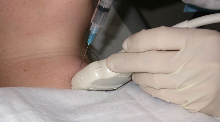
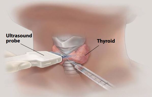
Your doctor will insert a small needle through the skin into the lump. Because FNA is about as painful as drawing blood from the arm for laboratory testing (venepuncture), local anaesthesia is usually not required. In fact, the needle used for FNA is smaller than that used for venepuncture. Although not painless, any discomfort associated with FNA is usually minimal. If the nodule is small or difficult to feel, ultrasound-guided FNA, performed by a specially trained radiologist, is used to help direct the needle into the mass.
The patient is instructed not to swallow or speak during the insertion of the needle. The aspiration may be done with the needle alone (moving to and fro while being rotated on its axis) or with a syringe attached to the needle. After the sampling, the needle will be removed. Samples of the cells are then put on a slide and fixed for review by the pathologist. The needle will be reinserted for additional samples. Several specimens are needed for a complete analysis. Once the FNA is complete, pressure will be applied to the area to decrease the risk of bleeding. A bandage is then placed.
This procedure is usually completed in less than 30 minutes.
WHAT WILL I EXPERIENCE DURING AND AFTER A FNA OF THE THYROID?
FNA of thyroid nodules are usually done under ultrasound guidance by a radiologist in the imaging department. During the test, you will lie on your back with a pillow under your shoulders, your head tipped backward, and your neck extended. This position makes it easier for the interventional radiologist to access the thyroid gland.
You may feel some pressure on your neck from the ultrasound transducer and mild discomfort as the needle is moved to obtain the cells. Limited intra-thyroidal bleeding and mild local pain radiating to the ear may occur. The most significant possible complication of the procedure is the development of a neck haematoma which may require a surgical intervention, but this complication is exceptionally rare. You will be charged for all extra treatments.
Generally, you can resume normal activities quickly and any bandage can be removed within 6 hours. The puncture site may be sore for 1 to 2 days. You may take paracetamol to relieve any discomfort.
HOW ABOUT THE RESULTS?
A pathologist in the laboratory will examine the removed specimen and will make a diagnosis. Tests for infection and certain chemical substances can also be done if required on the material that is obtained.
Results are usually given within a few working days.
There is a risk, because the samples aspirated are very small that the problematic cells will be missed, for example due to excessive blood or necrotic debris obscuring cellular details, resulting in a negative result. There is also a risk that the cells taken will not enable a definitive diagnosis. In those cases, your doctor will discuss with you the options to repeat the FNA or do a surgical biopsy.
For further information and for taking appointment, kindly contact our imagingthe Imaging department at (08028) 54 11 34 00