WHAT IS CONVENTIONAL ARTHROGRAPHY?
Conventional arthrography is the x-ray examination of a joint that uses a special form of x-ray called fluoroscopy and a contrast material containing iodine.
An x-ray (radiograph) is a non-invasive medical test that helps doctors diagnose and treat medical conditions. Imaging with x-rays involves exposing a part of the body to a small dose of ionizing radiation to produce pictures of the inside of the body. X-rays are the oldest and most frequently used form of medical imaging.
Fluoroscopy makes it possible to see internal organs in motion. When iodine is injected into the joint space, it coats the inner lining of the joint structures and appears bright white on an arthrogram, allowing the radiologist to assess the anatomy and function of the joint.
Today, most images are digital files that are stored electronically. These stored images are easily accessible and are sometimes compared to current x-ray images for diagnosis and disease management.
WHAT ARE SOME COMMON USES OF THE PROCEDURE?
Arthrographic images help doctors evaluate alterations in structure and function of a joint and help to determine the possible need for treatment, including surgery or joint replacement.
The procedure is most often used to identify abnormalities within the:
- Shoulder
- Wrist
- Hip
- Knee
- Ankle
The procedure is also used to help diagnose persistent, unexplained joint pain or discomfort.
HOW SHOULD I PREPARE?
No special preparation is necessary before arthrography. Food and fluid intake do not need to be restricted.
You will be asked to fill out a Questionnaire and a Consent form prior to the exam.
You should inform your doctor of any medications you are taking (written list of medications) and if you have any allergies, especially to iodinated contrast materials. Also inform your doctor about recent illnesses or other medical conditions.
You may be asked to remove some or all of your clothes and to wear a gown during the exam. You may also be asked to remove jewellery and any metal objects or clothing that might interfere with the x-ray transmission.
Women should always inform their doctor or the diagnostic radiographer if there is any possibility that they are pregnant. Many imaging tests are not performed during pregnancy so as not to expose the foetus to radiation. If an x-ray is necessary, precautions will be taken to minimize radiation exposure to the baby.
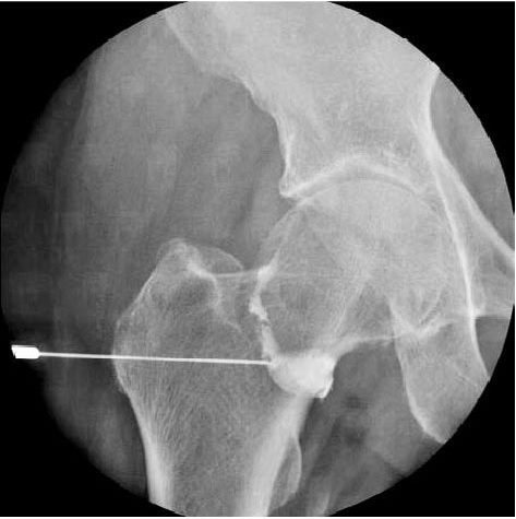
HOW IS THE PROCEDURE PERFORMED?
This examination is usually done on an outpatient basis.
The patient is positioned on the examination table and x-rays of the joint are taken to be compared later with the arthrograms.
Next, the skin around the joint is cleansed with antiseptic and in some cases; a local anaesthetic is injected into the area. A needle with an aspiration syringe is then inserted into the joint space. Next, the contrast material and – sometimes – air are injected into the joint space. You may feel a fullness as the joint is filled. Once the needle is removed, the patient will be asked to move the affected joint to distribute the contrast material throughout the space. Images are then obtained with the joint in various positions.
The examination is usually completed within 30 minutes.
CT scanner images may be obtained right after conventional arthrography to better evaluate the internal structures of a joint.
WHICH COMPLICATIONS MIGHT OCCUR?
Any interventional procedure performed on human being, even under maximal security conditions, maintains some risks of complication.
Because the contrast material is put in a joint and not a vein, allergic reactions are very rare, although in some cases, mild nausea to severe cardiovascular complications may result.
Like other puncture, there is a small risk of introducing an infection when a needle is placed in a joint. Your doctor will take all necessary precautions to avoid this. After the examination, you may experience swelling and discomfort. You may apply ice to the joint to reduce swelling. A mild over-the-counter analgesic can be taken for pain. These symptoms usually disappear after 48 hours. Contact your doctor if they persist after two days.
Vigorous exercise is not recommended for 24 – 48 hours after the exam.

HOW ABOUT THE RESULTS?
After the examination, the radiologist will give you some preliminary comments. He will, of course, analyse the images in details and a written report will be available as soon as possible.
A conventional arthrography has some limitations especially about the internal structures of a joint such as ligaments, meniscus, etc. That’s why nowadays; CT Arthroscan are frequently indicated in joint diseases.
HOW DO I SCHEDULE A CONVENTIONAL ARTHROGRAPHY ?