
What is general radiography (X-ray)?
A general radiography (X-ray) is an important diagnostic tool and the most basic form of diagnostic imaging. X-rays are used to create images of the body’s internal structures. These images are used to detect abnormalities in bones, lungs and other internal organs. X-ray involves exposing a part of the body to a small dose of ionising radiation to produce an image of the bones and internal organs to help better diagnosis and treat a patient’s condition. When X-rays penetrate the body, they are absorbed in varying amounts by different parts of the anatomy. Ribs and bones, for example, will absorb much of the radiation and, therefore, appear white or light gray on the image. Lung tissue and other internal organs absorb lesser radiation and appear darker on the image. In this manner a ‘picture’ of the body part is formed.
What are some common uses of general radiography?
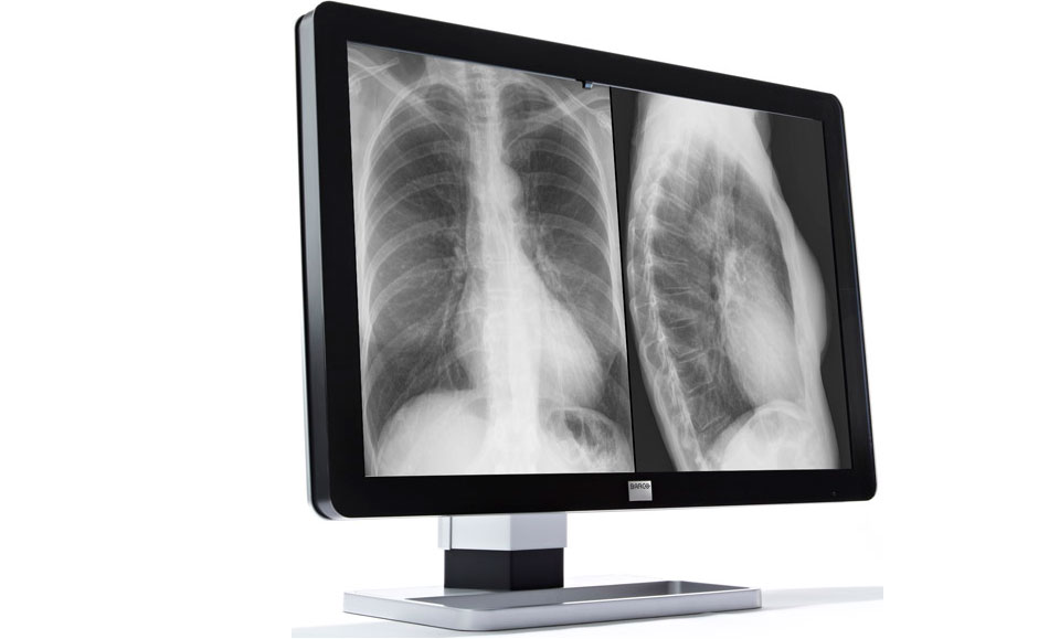
Chest and bone X-rays are very common examinations.
A chest X-ray is usually done for the evaluation of lungs, heart and surrounding anatomy.
Bone X-ray is the fastest and easiest way for a doctor to view and assess broken bones, cracked skull and injured backbone. Usually 2 images are taken of a bone, and often 3 or more if the problem is around a joint (knee, elbow or wrist).

What are the benefits and risks?
Benefits
X-ray imaging is useful to diagnose bone and joint injury and disease, such as fractures, infections, arthritis and cancer.
Because X-ray imaging is fast and easy, it is particularly useful in emergency diagnosis and treatment.
X-ray equipment is widely available, making it convenient for both patients and doctors.
Risks
Some people worry that X-rays aren’t safe because radiation exposure can cause cell mutations that may lead to cancer. The amount of radiation you’re exposed to during an X-ray depends on the tissue or organ being examined. Sensitivity to the radiation depends on your age, with children being more sensitive than adults.
Generally, however, radiation exposure from an X-ray is very low in modern imaging facilities using state-of-the-art equipment, and the benefit from these tests far outweighs the risks.
If you’re pregnant or you think you might be pregnant, tell your doctor or the radiographer before having an X-ray. Though the risk of most diagnostic X-rays to an unborn baby is small, your doctor may consider another imaging test, such as ultrasound. Generally, 10 days from the first day of the last menstrual period is considered safe period for X-ray examinations.
How should I prepare for the procedure?
The procedure requires no special preparation. You may be requested to change into a gown to avoid metallic items, buttons and zippers. You will also be asked to remove jewellery, eyeglasses and any metal objects that could obscure the image.
As X-rays are involved, it is not advisable for women who are pregnant, especially in early pregnancy. To prevent unnecessary irradiation of foetus, female patients will be asked about their pregnancy status prior to perform the X-ray. This is especially important in examination of the abdomen and pelvis.
How is the procedure performed?
A radiographer positions your body to obtain the necessary views. He or she may use pillows or sandbags to help you hold the position. Once you are positioned in the required pose with the X-ray detector, you may be asked to take a deep breath and hold it or just to hold your breath and keep still. The radiographer will go to another small room or cubicle and activate the X-ray equipment which will send a beam of X-rays to the positioned area. You need to keep still as any movement will lead to a blurred picture and an accurate diagnosis cannot be made.
X-ray exams are usually completed within 5 to 30 minutes depending on the number of views required by your doctor, the severity of your illness, your age and your co-operation.
When your X-ray exam is completed you will be asked to wait until the radiographer, and sometimes the radiologist, examines the images to determine if additional views are needed.
Your child’s X-ray
If your child is having an X-ray, restraints or other techniques may be used to keep him or her still. These techniques, which may be necessary if your child moves during the X-ray exposure, are safe, won’t harm your child and will prevent the need to repeat exposure to radiations. You may be asked to stay with your child during the test, and sometimes to help him to keep the good position. If you remain in the room during the X-ray exposure, you’ll be asked to wear a lead apron to shield you from unnecessary exposure. You must inform the radiographer if you’re pregnant or you think you might be pregnant.
What will I experience during the procedure?
This is a painless procedure. The only discomfort may result from the coldness and the hardness of the X-ray equipment or X-ray detector. Sometimes to get a clear image of an injury, you may be asked to hold onto an uncomfortable position for a short time. Any movement could blur the image and make it necessary to repeat the procedure to get a useful, clear picture.
When can I expect results?
X-rays are captured, distributed and stored in a digital format viewable on computer screens within minutes. A radiologist typically views and interprets the results and sends a report to your doctor, who then explains the results to you. In an emergency, your X-ray results can be made available to your doctor in minutes.
Related Articles
Related Articles

Virtual Colonoscopy (CT Colonography)
WHAT IS A VIRTUAL COLONOSCOPY? Virtual colonoscopy, also called CT colonography, is a method of screening the colon for precancerous polyps using a CT scanner. CT scanning is a non-invasive medical test that combines special x-ray equipment with sophisticated computers to produce multiple images of the inside of the body. CT colonography provides an interior […]
Percutaneous Radiofrequency Ablation of Liver Tumours
WHAT IS RADIOFREQUENCY ABLATION OF LIVER TUMOURS? Radiofrequency ablation (RFA) is a minimally invasive, repeatable procedure with few complications used to treat small, often inoperable, metastatic (particularly from colon cancer) and primary (hepatocellular carcinoma) tumours of the liver. Percutaneous RFA is an effective treatment option for patients who might have difficulty with surgery or those […]
Ultrasound
WHAT IS ULTRASOUND? Diagnostic ultrasound, also called sonography or diagnostic medical sonography, is an imaging method that uses high-frequency sound waves to produce images of structures within your body. The sound waves are recorded and displayed as a real-time visual image. No ionising radiation (X-ray) is involved in ultrasound scanning. In most ultrasound examinations, a […]
