
WHAT IS A MRI SCAN?
Magnetic resonance imaging (MRI) is a type of scan that is often used to diagnose conditions affecting organs, tendons and bones.
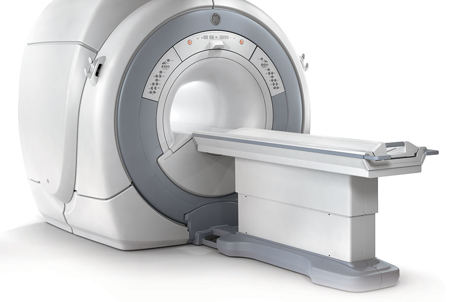
MRI scans use strong magnetic fields and radio waves to produce a detailed image of the inside of the body, particularly soft tissues, such as the brain, spinal cord and muscles. The device that performs MRI scans is known as a MRI scanner. The scanner consists of a large tube that contains a series of powerful magnets. The patient lies inside the tube during the scan. One of the main advantages of MRI is that, unlike X-rays, it does not expose the body to radiation. Though some discomfort may occur from having to lay still, MRI is otherwise a painless procedure and typically takes approximately 30-40 minutes to perform.
WHAT ARE THE BENEFITS OF A MRI SCAN?
The MRI system Optima™ MR360 1.5T, used at FV Hospital, combines the latest technology with a dedicated software to produce high-quality images in a short period of time. It is also one of the most patient-friendly MRI scanners, designed to ensure that patients feel comfortable and completely at ease.
This 1.5T MRI system provides non-interventional advances clinical application, such as:
- Musculoskeletal imaging, i.e. imaging of all the joints, tendons, bones and tissues of superior and inferior extremities such as the wrist, arm, elbow, hand, hip, thigh, knee, shin, ankle, and foot
- Neurological imaging, i.e. imaging of the skull, brain, cerebral vessels and spinal cord
- Head and neck imaging, i.e. imaging of the neck spine, ear cavity, orbit, nasal cavity, larynx, salivary glands, lymph nodes, pituitary gland
- Imaging of the chest wall, thoracic spine, thoracic aorta
- Lumbar spine imaging
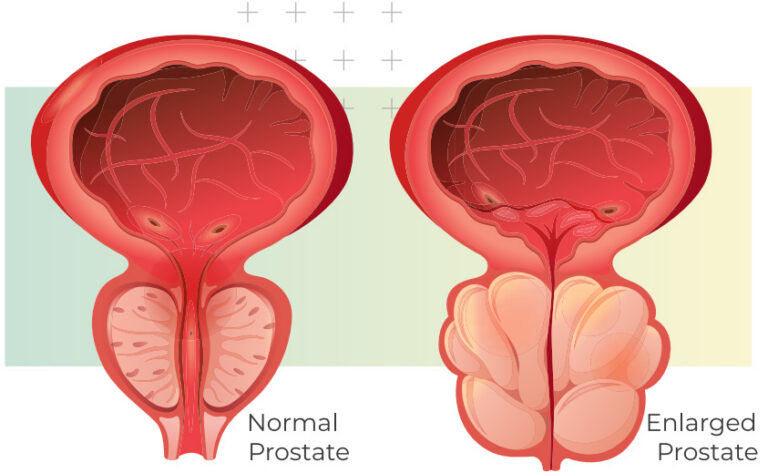
- Abdomen and pelvis imaging, i.e. imaging of the abdomen and abdominal wall, bile duct, pancreas, urinary system, uterus, cervix and ovaries (female), prostate and testicles (male), anal canal, pelvis floor, etc…
- Biliary and pancreatic duct imaging
- Vascular imaging (imaging carotid artery, cerebral blood vessels, aorta, internal arteries, extremity arteries)
- Breast imaging.
HOW SHOULD I PREPARE FOR THE PROCEDURE?
- No preparation is necessary for a MRI scan. You can eat and drink normally.
- On arrival at Imaging Department, you will be asked to complete MRI questionnaires before the scan to thoroughly understand your overall health and you will be required to sign a consent form for the MRI procedure.
- As a strong magnet is used, all metallic devices MUST be removed before entering the MRI room. Patients with cardiac pacemakers and cochlear implants cannot undergo MRI scans. Other metallic implants may prohibit patients from having a MRI scan. Therefore, you should inform the radiologists or the diagnostic radiographer if you have any of the following:
- Pacemaker
- Cerebral aneurysm clips, vascular stents, infusion pumps, neurostimulators
- Cochlear, otologic, or other ear implants
- Metal fragments in your eye
- Any other metal implants (surgical staples, artificial teeth, artificial joints, etc…)
- You must remove items like your jewellery, wallet, watch, keys, magnetic strip card (ATM, credit cards), hairpins, and phone.
- You will be asked to change into a gown to prevent metallic objects from being attracted by the powerful magnet. You will then lie on a scanning table that moves into the bore of the MRI. The body part to be scanned will be positioned in the centre of the tunnel. A device, known as a coil, which improves the quality of the images, may be placed over the region of interest.
- The scan occurs in an enclosed space, so if you have problems with this and may experience claustrophobia or anxiety, please inform us in advance.

Information for female patients:
Female patients are requested not to wear eye make-up for brain scans as this can affect image quality. If you are or might be pregnant, please inform staff of FV Hospital’s Imaging Department in advance of your appointment so that the radiologist can discuss your situation with yourself and/or referring doctor. As a general rule, MRI scans are usually not conducted in the first trimester of pregnancy unless it is deemed by the referring doctor or radiologist that is an absolute medical necessity to do so and that the benefits of the test outweigh the risks. This of course assumes that there is no other test available that can provide similar information.
WHAT WILL I EXPERIENCE DURING THE PROCEDURE?
- You will be positioned on a padded table and slowly moved into an open magnet that surrounds the body with a magnetic field.
- You will be given headphones so that you can listen to music if you want, which helps to reduce the whirring noise you hear from the MRI scanner. During the scan, the diagnostic radiographer will be able to talk to you through an intercom system.
- You will know when the scan is under way as you will hear a vibrating sound. It is very important that you keep your body extremely still. Movement will ruin the image produced, similar to the blurring effect that occurs when taking a photograph of a moving object.
- The radiologist and the diagnostic radiographer will review the images during the scan to make sure that they are clear.
MRI Contrast media (Gadolinium)
Some patients undergoing a MRI scan may require an injection of an intravenous contrast media known as Gadolinium, a substance that makes the lesion clearly visible on MRI scans. The contrast is delivered into your body through a small plastic tube known as an intravenous cannula, which is placed into a vein in your arm by a nurse or a diagnostic radiographer who are both experienced in performing this procedure. This will result in a minor discomfort, usually no more than taking blood from your arm. The contrast media is NOT radioactive.
The benefit of administering intravenous contrast media for a MRI examination is enormous. The use of contrast media greatly improves the accuracy of the examination and assists in excluding many life threatening conditions, such as cancer.
Most injections of IV contrast media occur uneventfully.

So that you are fully informed of the risks prior to the examination, FV Hospital’s Imaging Department would like to inform you that:
- As for all medical procedures, there are risks associated with the administration of any substance, including intravenous contrast media.
- If after reading the information below you are not willing to undergo a study with intravenous contrast media, the test may still be performed without it, however you must be aware that the information from the examination may not accurately answer your doctor’s question:
- When the contrast media is injected, it is normal to feel coolness and a flushing sensation for a minute or two. Some patients may sense a metallic taste in their mouth after the contrast media injection. A few patients experience side effects from the contrast media, including nausea and local pain. Very rarely, patients are allergic to the contrast media and experience hives, itchy eyes or other reactions. If you experience allergic symptoms, notify the diagnostic radiographer. A radiologist or other doctor will be available for immediate assistance. These minor reactions occur in less than 0.05% of cases. Our department is well-equipped to deal with them. There is no way of predicting who will be allergic to contrast media until the dye is given.
- Nephrogenic systemic fibrosis is currently a recognized, but rare, complication of MRI believed to be caused by the injection of high doses of gadolinium contrast media in patients with very poor kidney function.
Water-based gel contrast media:
Some patients undergoing a pelvic MRI scan may require a water-based gel filling of the vagina and the rectum that makes the lesion clearly visible on MRI scans. The gel is introduced into these cavities through a small plastic flexible tube by a diagnostic radiographer who is experienced in performing this procedure. This will result in a minor discomfort, and the gel can be easily released by going to the toilet after the procedure. For women, a sanitary napkin will be provided upon your request.
HOW LONG DOES A SCAN TAKE?
Usually four or five different types of MRI scans (sequences) are taken with each one lasting about 2-8 minutes. Overall, you will be in the scanner for about 30-40 minutes.
WHO INTERPRETS THE RESULTS AND HOW DO I GET THEM?
Your examination will be reviewed and reported by a radiologist. Your referring doctor will be informed immediately of any suspicious findings. You will receive a formal written report on the same day or within two (2) days depending on your clinical and diagnostic requirements.
Please ensure that you make a follow up appointment with your referring doctor or health care provider to discuss your results.
HOW DO I SCHEDULE THE SCAN?
FOR FURTHER INFORMATION AND APPOINTMENTS, KINDLY CONTACT OUR IMAGING DEPARTMENT AT (08) 54 11 34 00
REMEMBER
- Please bring to the hospital the prescription from your doctor for the MRI, any prior scans (i.e. X-rays, ultrasounds, MRI, CT) and reports as these will assist the radiologist in assessing your condition.
- Please note that any referral for a scan is valid at Imaging Department of FV Hospital, even if it has been written on a referral form from another radiology provider.
- Whilst every effort is made to keep your appointment time, the special needs of complex cases, elderly and frail patients can cause unexpected delays. Your consideration and patience in these circumstances is appreciated.
Related Articles
Related Articles

Virtual Colonoscopy (CT Colonography)
WHAT IS A VIRTUAL COLONOSCOPY? Virtual colonoscopy, also called CT colonography, is a method of screening the colon for precancerous polyps using a CT scanner. CT scanning is a non-invasive medical test that combines special x-ray equipment with sophisticated computers to produce multiple images of the inside of the body. CT colonography provides an interior […]
Percutaneous Radiofrequency Ablation of Liver Tumours
WHAT IS RADIOFREQUENCY ABLATION OF LIVER TUMOURS? Radiofrequency ablation (RFA) is a minimally invasive, repeatable procedure with few complications used to treat small, often inoperable, metastatic (particularly from colon cancer) and primary (hepatocellular carcinoma) tumours of the liver. Percutaneous RFA is an effective treatment option for patients who might have difficulty with surgery or those […]
Ultrasound
WHAT IS ULTRASOUND? Diagnostic ultrasound, also called sonography or diagnostic medical sonography, is an imaging method that uses high-frequency sound waves to produce images of structures within your body. The sound waves are recorded and displayed as a real-time visual image. No ionising radiation (X-ray) is involved in ultrasound scanning. In most ultrasound examinations, a […]
