From October 13th to 15th, 2023, a scientific conference titled “Updates in the Management of Congenital Heart Disease and Structural Heart Disease: Foetal to Adult Patients” was held in Ho Chi Minh City. The conference was organised by the Vietnam Echocardiography Society. During the conference, Dr Nguyen Van Te, a leading expert at FV Hospital in the field of nuclear medicine, provided updates on new knowledge related to diagnosing the heart condition amyloidosis using nuclear imaging techniques.
A rare condition due to underdiagnoses
Amyloidosis, or cardiac amyloidosis, is a rare condition characterised by the abnormal deposition of insoluble protein fibres in the extracellular space, causing damage to the heart. This is a rare and highly dangerous condition that has been underdiagnosed until recently that continues to pose challenges in the fields of medical diagnosis and treatment. In addition to affecting the heart, amyloidosis can also damage the nervous system, respiratory system, digestive system, liver, kidneys, and the thyroid gland, among other areas.
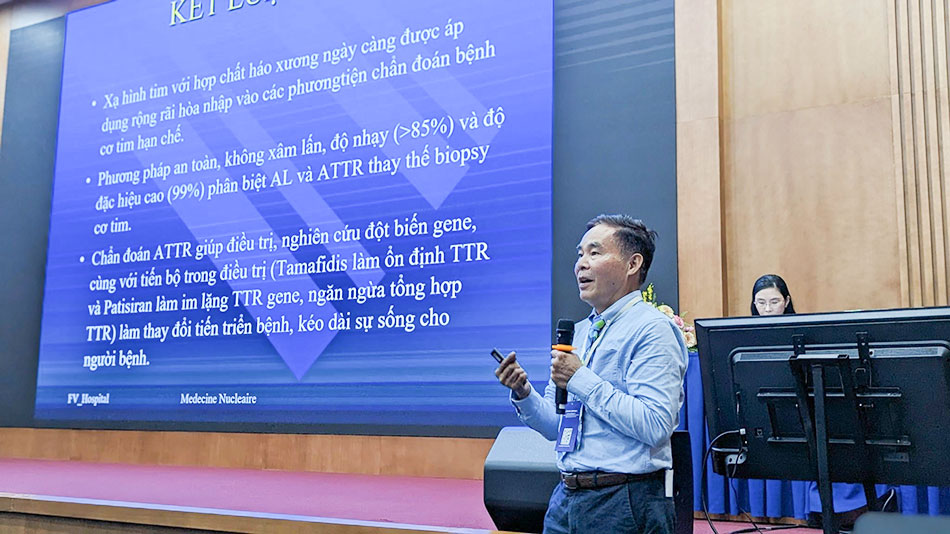
Dr Nguyen Van Te, Head of Nuclear Medicine Department, FV Hospital, presents at the conference.
In his report on “Nuclear Imaging in Diagnosis of Amyloid Cardiomyopathy,” Dr Nguyen Van Te, Head of the Nuclear Medicine Department at FV Hospital, indicates that the causes of cardiac amyloidosis can be divided into two main groups.
The first is amyloidosis originating from disorders in plasma cell-produced light chain immunoglobulins (AL), which leads to excessive light chain immunoglobulin (Ig) production. This form accounts for approximately 2500 new cases each year. From the time of diagnosis with heart failure symptoms, the life expectancy of these patients is usually less than one year.
The second group involves transthyretin-related amyloidosis (ATTR), a type of protein synthesised in the liver with a misfolded process. ATTR can be further categorised into hereditary (hATTR) when a gene mutation is present or wild-type (wATTR) ATTR, typically seen in people over 60 years of age. ATTR-related amyloidosis tends to progress slowly, is difficult to diagnose, and often remains asymptomatic until serious cardiac complications arise. Without early treatment, the remaining life expectancy for affected individuals is approximately 10 years.
Currently, non-invasive methods such as blood and urine tests, electrocardiograms, echocardiography, and cardiac MRI aid in diagnosing cardiac amyloidosis. However, there are still significant limitations and uncertainties in the diagnosis. As a result, cardiac hypertrophy due to amyloid infiltration remains a hot topic at specialised heart failure conferences worldwide.
Nuclear Imaging should be more widely disseminated for the diagnosis of cardiac amyloidosis.
To accurately diagnose cardiac amyloidosis, heart biopsy is considered the gold standard. However, this is an invasive procedure associated with potential complications and not widely available.
“Nuclear imaging is a safe, non-invasive method with a sensitivity of over 85 per cent and high specificity of up to 99 per cent in distinguishing AL and ATTR. This method can completely replace heart biopsy, it is cost-effective, and patients do not need the same preparations as they would for surgery,” emphasised Dr Te.
However, nuclear imaging has not been widely adopted in many places due to equipment costs and is not yet widely used in Vietnam or worldwide for cardiovascular disease diagnostics.
Dr Nguyen Van Te described how a special bone-seeking radiopharmaceutical molecule called Technetium-99m is injected intravenously before the patient is scanned using single photon emission computed tomography/computed tomography (SPECT/CT). The result is anatomical images along with quantitative and qualitative assessment of cardiac radiotracer uptake, allowing doctors to accurately determine the patient’s cardiac pathological condition.
The protocol that Dr Te has implemented at FV is based on the American Society of Nuclear Cardiology (ASNC) guidelines. Additionally, Dr. Te proposed four diagnostic scenarios for this condition according to the new guidelines of the European Society of Cardiology: combining scintigraphy, assessing monoclonal proteins, performing cardiac magnetic resonance (CMR), and heart biopsy.
Dr Te also presented several cases of cardiac amyloidosis that were accurately diagnosed at the Cardiology and Nuclear Medicine Department at FV for the first time in February 2021. Along with rare, disease-specific drugs newly approved by the FDA, the use of scintigraphy technology in diagnosing cardiac amyloidosis, especially in detecting the disease in its early stages when symptoms are still vague, has provided patients with the opportunity for effective treatment, disease management, and screening for family members.
The Nuclear Medicine Department is an FV specialty that has operated since the early days of the Hospital, providing imaging diagnostics through Gamma cameras for SPECT/CT scans, PET scans, etc. which can replace previous exploratory surgeries. Nuclear scanning is useful for diagnosing coronary artery diseases, thyroid disorders, determining pulmonary embolism, evaluating renal morphology and function, diagnosing adrenal gland tumours, and metastatic bone cancer assessment, among other functions.