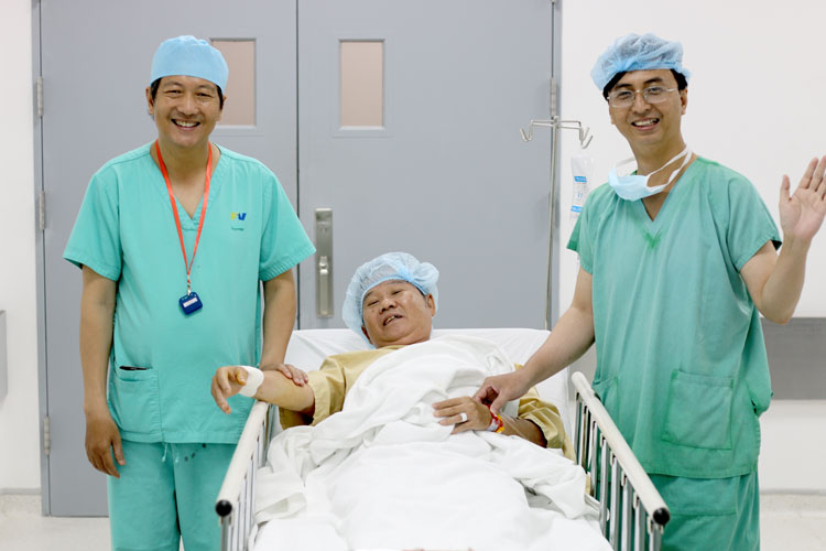
On July 4, 2018, FV Hospital docotors successfully performed an autolytic stent placement in combination with intravascular ultrasound for patient Mr Mai Buu Ngoc, who was experiencing stenosis (narrowing) of his coronary artery. This is an advanced technique to minimise risks and complications and prevent the renarrowing and blockage of blood vessels due to thrombosis.
Every morning, Mr Ngoc, who resides in District 7, HCMC, experienced shortness of breath and pain in the left side of his chest. The frequency and extent of his symptoms increased over time. Although Mr Ngoc was taking medication, his symptoms did not improve. He decided to go to FV Hospital to be examined by doctors. Initial diagnostics showed that Mr Ngoc’s coronary artery network had narrowed by 80 per cent in the pre-interventricular artery and 70 per cent in the arterial branch, and required an angioplasty – the surgical repair or unblocking of a blood vessel. However, at age 53, Mr Ngoc was very worried about his prognosis and having surgery related to such an important part of the body as the heart.
Understanding Mr Ngoc’s concerns, Dr Huynh Ngoc Long, head of Cardiology at FV Hospital, explained the details of the procedure to his patient, using illustrations to show stages of the surgery, introducing potential complications and their solutions, and finally outlining expected results. After thorough consideration, Mr. Ngoc decided to undergo surgery.
According to the patient’s preferences, FV Hospital doctors chose to place autolytic Magmaris-Biotronik stents, made in Switzerland and used in many European countries, into the patient’s blood vessels. This autolytic stent frame is made of Magnesium alloy with just a small concentration of metal so that it can dissolve in the body around 12 months after being placed.
In order to accurately assess the degree of blockage in the patient’s intravascular blood vessels, FV’s cardiology surgeons used intravascular ultrasound (IVUS). With precise support from this device they were enabled to examine the blockage in situ and could confidently choose the right stent and hard ball size to use.
Specifically for Mr Ngoc, in the pre-interventricular artery, cardiologists used a 3.0 mm intravenous catheter for a 3.5 mm intraocular vein, and selected a 3.5 mm stent and performed a 3.75 mm angioplasty after stent placement.
In the arterial branch, the narrowed blood vessel measured 2.5 mm in diameter, while the actual IVUS showed a 3.3 mm wide blood vessel. Doctors selected a 3.0 mm stent and a 3.5 mm angioplasty following stent placement. By selecting the optimal stent, the diameter of the intravascular blood vessels can be maximised and the stent could better fitted to the blood vessel.
After nearly two hours of implementation, the first autolytic stent placement combined with intravascular ultrasound at FV Hospital was successful. Mr Ngoc remained awake throughout surgery as the surgeons had not needed to give him a general anaesthetic.
Mr. Ngoc said:
Satisfied with the success of the surgery, Dr Huynh Ngoc Long added: “Compared to the drug-coated stent, which has a metal frame, the autolytic stent has many benefits for patients as it can dissolve in the body a year after being placed. After stent placement, blood vessel elasticity is restored and bridging surgery can be performed. A great benefit for patients is that chest pain after the procedure is minimised.”
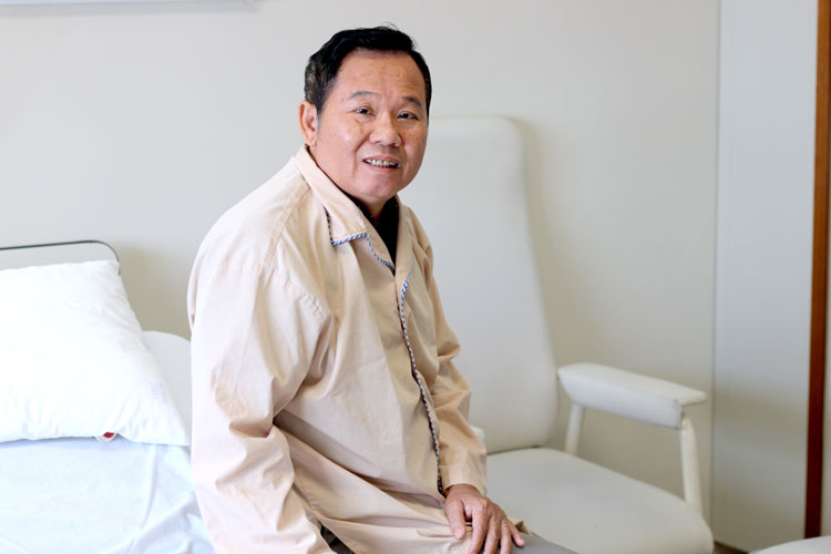
After a day of rest at FV Hospital, Mr. Ngoc was completely recovered and could be discharged from the hospital.
The use of autolytic stent placement combined with intravascular ultrasound is an extremely effective treatment option for cardiovascular patients. Although there are certain considerations, such as the age of the patient, stent size limitation, the patient taking anticoagulants for at least six months, dilation with hard shade 1:1, and increased pressure in the blood vessels treated, the benefits are considerable: the patient’s blood vessels are clear, with restored elasticity and a reduced risk of thrombosis.
By performing more procedures like this, FV Hospital will contribute to the development of diagnostic technology and the direction of treatment for coronary artery disease. As a result, doctors can offer more effective treatments to prevent serious and dangerous complications, reducing the cost of long-term treatment for patients.
FV’s Cathlab is operated by a team of experienced doctors, led by Head of Cardiology Dr Huynh Ngoc Long, who has more than 22 years of experience. He is ranked among the top three cardiology doctors in Vietnam and specialises in the treatment of coronary heart disease, congenital heart disease, and other heart valve diseases.
To make a cardiology appointment at FV Hospital, please contact 028 5411 3467 (extension 1216 or 1165).