Four years ago, Mrs T.T.M.L. (56 years old, Ho Chi Minh City) started to experience symptoms of tiredness and her chest feeling tight, like a drum. These signs rarely appeared, but when they did, they lasted around 10 minutes before her condition returned to normal. For this reason, Mrs T.T.M.L. did not pay too much attention to her symptoms.
After several months, she was indicated for urethral augmentation with a TOT at the Urology Department, FV Hospital. During the pre-anaesthesia examination, the doctor found that Mrs T.T.M.L.’s heartbeat had an abnormal rhythm and so she was unable to undergo surgery. She was transferred to the Cardiology Department for a more in-depth diagnosis. There, Mrs T.T.M.L. was diagnosed with premature ventricular contractions, a heart rhythm disorder caused by abnormal electrical impulses in the heart muscle. The doctor advised that she be treated with electrophysiological ablation so that Mrs T.T.M.L. could receive her planned surgery soon while avoiding the risk of potential complications such as heart failure and sudden death.
Mrs T.T.M.L. shares: “I felt that my disease was not serious, and that I could delay treatment without any risk.”
With her wishes in mind, doctors prescribed a course of medicine as a temporary treatment. After three years of medication, the drugs became less effective and Mrs T.T.M.L.’s symptoms of fatigue began to appear more regularly and last longer. Mrs T.T.M.L. checked in with her doctor and decided to treat her condition completely by electrophysiological ablation.
This electrosurgery procedure is performed in FV’s Interventional Catheterisation Laboratory (Cathlab) with specialist equipment, including a three-dimensional electro anatomy imaging machine (3D mapping), which allows doctors to comprehensively treat premature ventricular contractions much more effectively than via internal medicine. Doctors perform local anaesthesia around the patient’s thigh vein to establish the entry route, then painlessly insert the catheter intravenously to the heart, moving it in the heart chamber to obtain a three-dimensional image and marking the suspected location using colour-coded electrical signals. Then, doctors isolate the ventricular external systole via radiofrequency therapy.
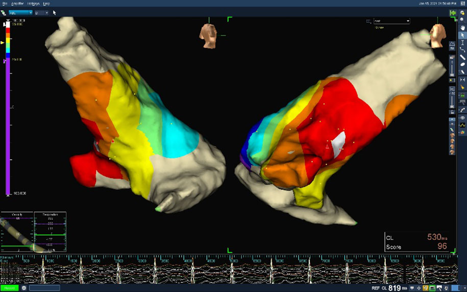
Dr Hoang Quang Minh, FV Hospital’s Cardiology Department, performed this procedure for Mrs T.T.M.L. and explains: “3D mapping uses fewer X-rays, which means fewer side effects compared to 2D image technology. It also helps us to identify the ventricular external systole more quickly.”
Mrs T.T.M.L.’s electrophysiological ablation took around two hours to complete. Her heart was simulated through 3D mapping, which helps doctors to locate the ventricular external systole more accurately. Combined with reports of interference shown in different colours, doctors burn the abnormal ventricular external systole preciously and quickly, avoiding any burning of the surrounding tissue, as well as repeated burning in a single location, which carries the risk of dangerous complications such as perforation of the heart and pericardial effusion.
Due to problems related to her heart, when Mrs T.T.M.L. requested her doctors give her general anaesthesia they advised her that it wasn’t suitable for her, and also that it wasn’t necessary.
“Dr Minh said this method is simple and fast, with few complications, and the team should not use general anaesthesia,” she shared.
After one month under undergoing treatment for premature ventricular contractions with radiofrequency therapy, Mrs. T.T.M.L. no longer needed to take medication. “I feel 98 per cent better,” she said happily.

FV Hospital’s Cardiology Department is equipped with state-of-the-art synchronic tools and technologies and is staffed by a highly experienced team of doctors. The Cardiology team is always ready to provide advanced diagnostic and comprehensive treatment plans, including prevention methodologies, early screening and diagnosis, treatment and cardiac rehabilitation.
Since May 2018, FV Hospital has operated its Interventional Catheterisation Laboratory (Cathlab). The Interventional Catheterisation Laboratory (Cathlab) received more than US$ 1.6 million of investment for its system of modern machines and equipment to best aid doctors with diagnosis and treatment of dangerous diseases. The conditions treated here include: heart attacks, blockages of blood vessels, aortic aneurysm and valve incompetence.
Dr Hoang Quang Minh is a senior doctor at the Cardiology Department, FV Hospital. Dr Minh has more than 10 years of experience in the treatment of cardiac arrhythmias with electrophysiological methods, ablation and pacing.
To make an appointment and to learn more, please contact the Cardiology Department: (028) 5411 3467 or (028) 35 11 33 33 Ext: 1216 / 1165.