
State-of-the-Art Technology
We use advanced imaging technologies, including high resolution ultrasound, 3D mammography, contrast imaging (CEM, MRI). Each modality is applied appropriately and combined when needed to improve diagnostic accuracy and provide a clearer understanding of each condition.
Advanced technology plays an important role in modern breast care but it is not about using more, it is about using the right tools in the right way.
We utilise a range of imaging technologies, including high-resolution ultrasound, 3D mammography (tomosynthesis), and contrast-enhanced imaging when appropriate. Each modality provides a different layer of information, helping us see breast tissue more clearly and evaluate findings with greater confidence.
These technologies are not used routinely for everyone. Instead, they are selected based on your individual situation and combined when needed to improve diagnostic accuracy. At FV Hospital, technology is guided by clinical judgment, supports decision-making but does not replace clinical expertise. Each test is chosen with a clear purpose, based on your individual needs.
The goal is simple: to provide a clearer, more complete understanding of each condition so that decisions can be made with confidence.
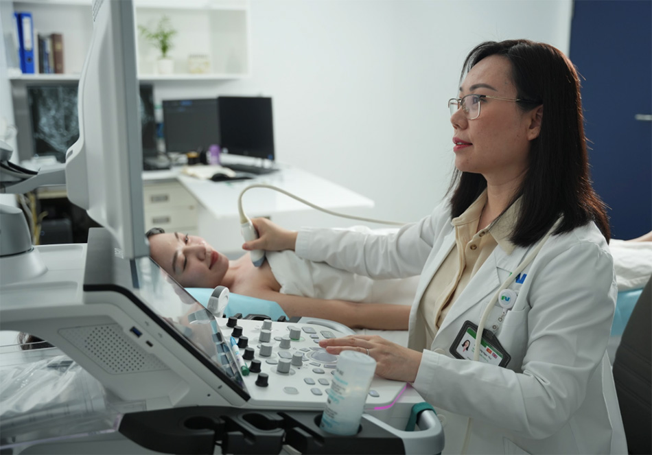
Ultrasound provides real-time, detailed evaluation of breast tissue, especially useful in dense breasts and for assessing specific areas of concern. Ultrasound is often used as a supplemental breast cancer screening tool, following a mammogram, to get additional information necessary for a complete diagnosis. Ultrasound provides real-time imaging, which may be used in guiding procedures such as biopsies. Ultrasound can help a radiologist determine whether a lump or abnormality is fluid-filled, fatty, or a solid mass.
Looking for expert breast care?
Contact us for consultation and appointment booking
Make an Appointment