

The Da Vinci Xi robotic system, integrated with ICG fluorescence imaging, is enabling greater precision in early-stage lung cancer surgery at FV Hospital. By clearly identifying anatomical segment boundaries during surgery, FV surgeons successfully removed an 11 mm lung tumour in a 68-year-old woman while preserving nearly 90% of her healthy lung tissue.
An 11 mm lung nodule discovered during a routine health check-up
Mrs N.D.T.N. (1958, Dak Lak Province) discovered an 11 mm lung nodule during a routine health check-up. As she had no cough, no fever and was able to carry on with her daily activities as usual, she could hardly believe the diagnosis when she received her results.
Concerned about the diagnosis, she sought consultation from Dr Dang Dinh Minh Thanh, PhD, Specialist Level II – Head of the Thoracic Surgery Department and the FV da Vinci Robotic Surgery Centre.
The CT scan revealed an 11 mm nodule in the lower lobe of the left lung, suggesting Stage 1A lung cancer. Dr Thanh ordered further scans of the head and abdomen, which showed no other lesions. According to international medical guidelines, surgical removal of the tumour is considered the preferred treatment option in cases like this, making a biopsy unnecessary.
“Fortunately, Mrs N.’s nodule was detected at an early stage while it was still small. She does not require removal of the entire lung lobe, removing only around 10% of her left lung would be sufficient to remove the tumour completely.,” explained Dr Dang Dinh Minh Thanh.
After receiving detailed counselling about the benefits of robotic surgery, including less pain, fewer complications, less blood loss and faster recovery, the patient and her family decided to proceed with surgery using the Da Vinci Xi robotic system. “Dr Thanh thoroughly explained the advantages of robotic surgery. After hearing his explanation, I agreed to the procedure straight away because I trust his expertise,” Mrs N. shared.

Dr Dang Dinh Minh Thanh, PhD, Specialist Level II, explains the Da Vinci Xi robotic system to Mrs N. and her family. Photo: FV
Robotic lung tumour removal preserves nearly 90% of healthy lung tissue with ICG technology
Lung segmentectomy is a highly specialised procedure requiring a high level of precision, as the boundaries between lung segments are difficult to distinguish during surgery. In Mrs N.’s case, the procedure was even more challenging because the nodule was located close to the border of the upper lobe of the left lung.
According to Dr Dang Dinh Minh Thanh, before advanced technology became available, surgeons often had to perform a larger resection to ensure the lesion was completely removed.
With the Da Vinci Xi robotic system, this challenge can be overcome using integrated Indocyanine Green (ICG) fluorescence imaging. Once injected into the patient’s body, the fluorescent agent creates a clear contrast between the tissue to be removed and the surrounding healthy tissue.
During Mrs N.’s operation, after removing the tumour by dividing the blood vessels and bronchus of left segment 6 and performing lymph node dissection, the surgical team switched the 3D monitor to fluorescence mode. The target lung segment was appeared clearly in green on the screen in real time, allowing the surgeon to clearly identify the surgical borders before making the resection.
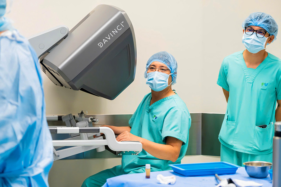
Dr Dang Dinh Minh Thanh, PhD, Specialist Level II, controls the robotic arms during tumour removal and lymph node dissection. Photo: FV
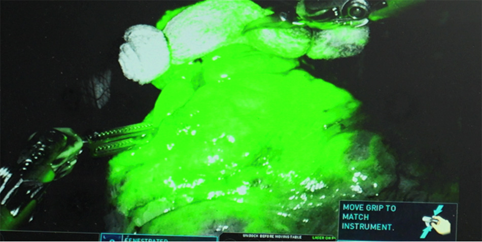
ICG technology helps visualise lung segment boundaries using fluorescence imaging. Photo: FV
After approximately one hour of surgery, the team successfully removed the 11 mm lung nodule, removed the relevant lymph node groups, and preserved nearly 90% of the patient’s healthy lung tissue. Mrs N. recovered well and was discharged two days after surgery.
“I would like to thank Dr Thanh for his advice and for treating me successfully. The nurses took excellent care of me. Even at night, whenever I needed help, they came straight away and were always cheerful, kind and attentive,” Mrs N. shared on the day of her discharge.
ICG Technology Expands the Possibilities of Precision Cancer Surgery
An increasing number of patients at FV Hospital are choosing robotic surgery for cancer treatment because of its precision and minimally invasive approach. With the Da Vinci Xi system, surgeons can operate more precisely in confined spaces and access deep or complex areas that may be difficult to reach with conventional laparoscopy. This allows more thorough tumour removal and lymph node dissection while minimising damage to surrounding healthy tissue.
Compared with open surgery, robotic surgery uses small incisions of around 8 mm, helping to reduce blood loss, minimise post-operative pain and shorten recovery time. The conversion rate to open surgery is also very low, at less than 1%.
Particularly in chest and gastrointestinal surgery, the integrated ICG fluorescence technology on the Da Vinci Xi system helps surgeons assess tissue and blood vessels in real time during surgery. In gastrointestinal procedures, ICG helps assess blood flow at the anastomosis site, reducing the risk of poor blood supply that may lead to leaks or delayed healing, among the most concerning post-operative complications.
To help patients access this advanced technology, FV Hospital is offering a discount of VND 40 million for the first 50 Da Vinci Xi robotic surgeries, valid until 15 June 2026. For more information, please contact FV Hospital at 6 Nguyen Luong Bang St., Tan My Ward, Ho Chi Minh City, Tel: (028) 3511 3333.
