



Mrs Chev Cheng, a 54-year-old Cambodian national, was diagnosed with lung cancer during a routine health check. Owing to early detection, she underwent successful robotic-assisted surgery using the da Vinci Xi system at FV Hospital, negating the requirement for further chemotherapy or radiotherapy.
A pulmonary nodule growing in silence
During a routine health check, Mrs Cheng chose to include low-dose CT lung cancer screening. The results revealed a pulmonary nodule of approximately 7mm. Because the lesion was still very small, her doctors scheduled a follow-up examination three months later to monitor its development. At the next check-up, the nodule had grown to approximately 15mm, doubling in size. Concerned by this development, Mrs Cheng’s daughter made the decision to bring her mother from Cambodia to FV Hospital for further evaluation and treatment.
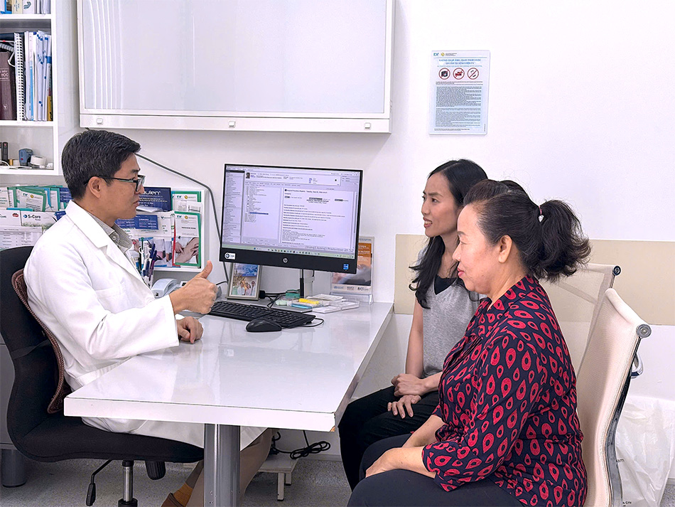
Dr Luong Ngoc Trung, MSc, Specialist Level II, explaining the patient’s health condition to her. Image: FV
At FV Hospital, doctors performed a CT-guided percutaneous transthoracic needle biopsy, a minimally invasive procedure that uses a needle to obtain a tissue sample from the lesion through the chest wall, avoiding open surgery. The result confirmed a diagnosis of non-small cell lung cancer.
Dr Luong Ngoc Trung, MSc, Specialist Level II, robotic surgery specialist and Head of the Vascular and Endovascular Surgery Department at FV Hospital, commented: “Mrs Cheng is a fairly typical presentation of early-stage lung cancer: a non-smoker, with no family history of the disease and no symptoms whatsoever. Without low-dose CT screening, it would have been extremely difficult to detect this tumour whilst it remained amenable to curative treatment.”
After a thorough assessment, Dr Trung confirmed the tumour was localised within the lung with no evidence of metastasis. Mrs Cheng’s case was presented to the Multidisciplinary Tumour Board, where thoracic surgery specialists, oncologists, and radiologists reached a consensus that robotic surgery using the da Vinci Xi system was the most effective treatment approach.
Early surgery, no adjuvant chemotherapy required
On June 2, Dr Trung and his team successfully performed a robotic pulmonary lobectomy with mediastinal lymph node dissection, completing the procedure in approximately 120 minutes.
Thanks to the high-definition 3D magnification and the ability to manoeuvre with exceptional precision within the confined space of the thoracic cavity, robotic surgery minimises injury to healthy surrounding tissue, reduces post-operative pain, and supports a faster recovery.
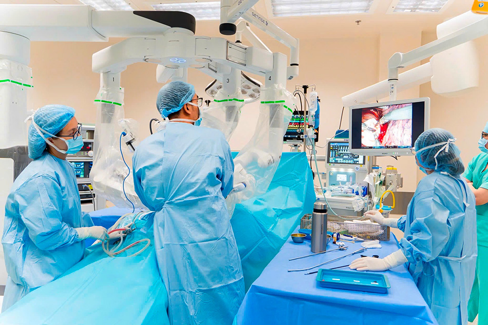
Robotic lung cancer surgery using the da Vinci Xi system. Image: FV
One day after the procedure, Mrs Cheng was walking independently, eating normally, and resuming her daily activities almost as before. Following a three-night observation period, she was discharged in stable condition.
“I am deeply grateful for the dedication of the doctors and nurses at FV Hospital. Their constant encouragement and attentiveness made me feel truly reassured throughout my treatment journey,” Mrs Cheng said.
Post-operative pathology results brought welcome news for the family. Histopathological analysis confirmed stage IA non-small cell lung cancer, with complete surgical resection achieved.
According to Dr Luong Ngoc Trung, patients with stage IA who undergo curative-intent surgery with complete tumour resection typically require only regular surveillance imaging thereafter, with no need for adjuvant chemotherapy or radiotherapy. Proactive screening had spared both the patient and her family not only a serious health risk, but also the enormous financial and emotional burden that advanced cancer treatment can place on a family. One that can, in some cases, prove completely overwhelming.
“What I am most relieved about is that my mother didn’t have to go through rounds of prolonged treatment. After just one operation, she was able to return to normal life,” said Kim Sovannavy, Mrs Cheng’s daughter.
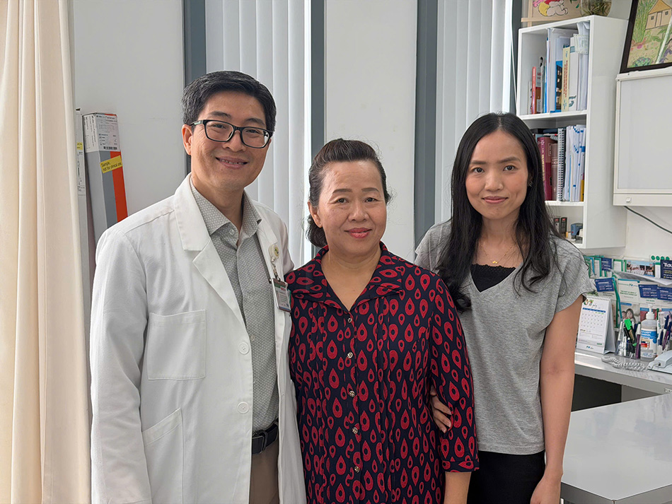
Mrs Cheng is fully recovered during her follow-up appointment. Image: FV
Why do experts recommend low-dose CT screening for lung cancer?
According to Dr Luong Ngoc Trung, MSc, Specialist Level II, the vast majority of patients are diagnosed only when the disease has already reached an advanced stage, presenting with symptoms such as a persistent cough, unexplained weight loss, chest pain, or signs of metastasis. At this stage, surgery is less effective, and patients often require a combination of chemotherapy, radiotherapy, or other systemic therapies at significantly higher cost.
“When lung cancer is detected at the earliest stage, five-year survival rates can reach approximately 90–95% in selected surgical series. This falls to approximately 53–65% at stage II, around 13–36% at stage III, and below 10–15% at stage IV,” Dr Trung said.
For this reason, more countries now recommend low-dose CT lung cancer screening for adults aged 50 and over, including non-smokers and those without clear risk factors.
Low-dose CT uses approximately 1–1.5 mSv) of radiation, roughly half the average annual background radiation dose to which we are all naturally exposed, and approximately 1/5 of the dose delivered by a standard chest CT, yet it remains sensitive enough to detect pulmonary nodules just a few millimetres in diameter. Chest X-ray, by contrast, can miss small lesions and is no longer recommended as a primary method for lung cancer screening by current international guidelines. For information about lung cancer screening programmes and robotic lung cancer surgery at FV Hospital, please visit us at 6 Nguyen Luong Bang Street, Tan My Ward (formerly District 7), Ho Chi Minh City, or call (028) 3511 3333. FV Hospital coordinates Social Health Insurance benefits, private health insurance, and 0% interest instalment payment plans to support patients with the financial aspects of cancer treatment and other conditions.
Related News
See moreRelated News
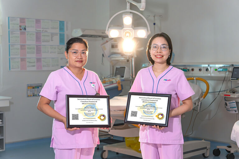
Two Vietnamese midwives attain international IBCLC certification in breastfeeding counselling
Two midwives from FV Thomson’s Maternity Department have earned IBCLC (International Board Certified Lactation Consultant) certification, the international credential for breastfeeding support specialists. This achievement represents a significant clinical milestone in infant feeding support at FV Hospital, driven by the real-world complexities these practitioners encounter daily when supporting mothers and their newborns. Beyond certification: A […]
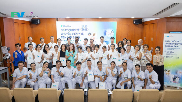
FV Hospital Celebrates International Allied Health Professions Day 2026
On 14 October 2026, FV Hospital held a special event to express its appreciation for allied health professionals. Attendees included members of the Board of Directors, heads of departments, and more than 40 allied health professionals and specialists. The event recognised the dedication of allied health professionals across a range of specialties, including Dietetics and […]
FV pioneers ERAS® implementation in Vietnam in line with international standards, supporting faster postoperative recovery
FV Hospital has become the first hospital in Vietnam to partner with Sweden-based Encare AB to implement the Enhanced Recovery After Surgery (ERAS®) programme. Through this partnership, FV joins Encare’s network of 22 hospitals in 11 countries across the Asia-Pacific region. The initiative aims to standardise surgical care according to international guidelines, leverage data to […]

Nutcracker Syndrome: How a one-hour stent placement ended a year of abdominal pain
FV Hospital successfully treated a complex case of nutcracker syndrome using minimally invasive endovascular stenting, sparing the patient major open surgery. Persistent abdominal pain linked to renal vein entrapment For over a year, 41-year-old Ms V.T.K.P. from Ho Chi Minh City experienced persistent, dull lower abdominal pain that worsened with prolonged sitting or standing. As […]
Two Vietnamese midwives attain international IBCLC certification in breastfeeding counselling
FV Hospital Celebrates International Allied Health Professions Day 2026
FV pioneers ERAS® implementation in Vietnam in line with international standards, supporting faster postoperative recovery

