
Early Detection of Breast Cancer
Early detection improves outcomes and reduces the need for complex treatment. Screening is tailored based on age, risk factors, and clinical context, using appropriate imaging methods to detect abnormalities early and guide timely, effective care.
Breast cancer is the second leading cause of cancer-related deaths in women. Early detection can make a significant difference in breast cancer care. When abnormalities are identified at an earlier stage, treatment is often more effective and may be less extensive.
Screening is not the same for everyone. The right approach depends on age, breast density, family history, personal risk factors, and clinical findings. For this reason, screening should be tailored rather than applied as a fixed routine.
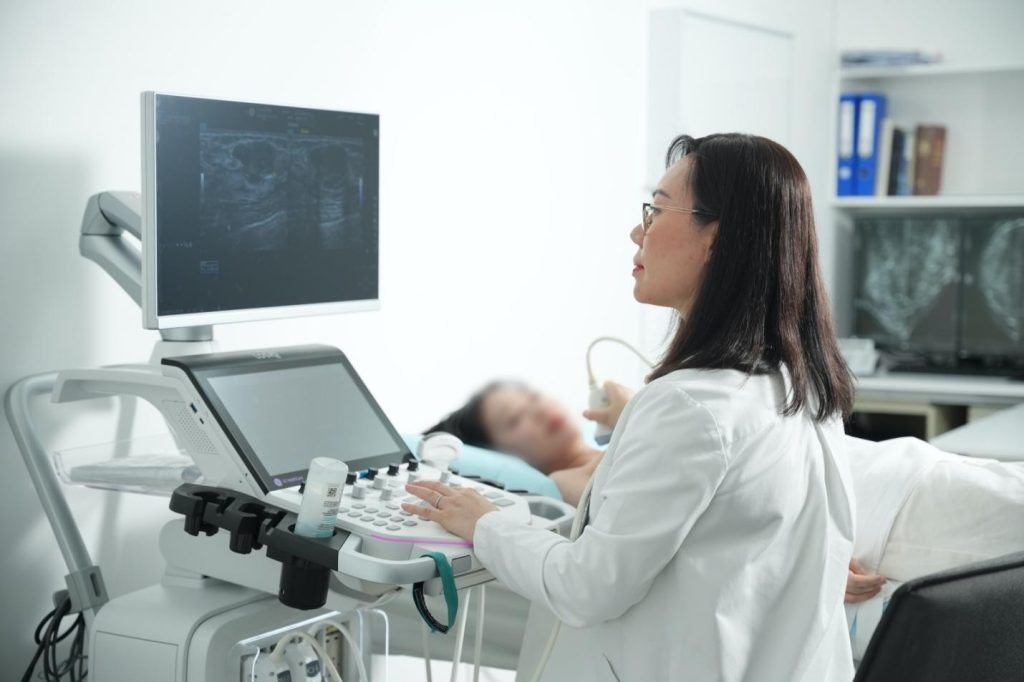
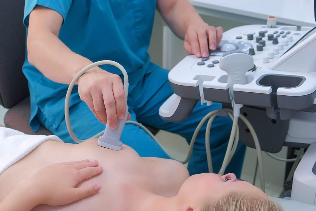
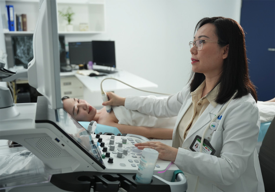
Different imaging methods may be used depending on the situation, including ultrasound, mammography, or MRI in selected cases. The aim is to detect meaningful abnormalities as early as possible and guide timely, appropriate care.
Early detection is not simply about finding more. It is about finding what matters early enough to make a real difference.
Tailored Screening Approach

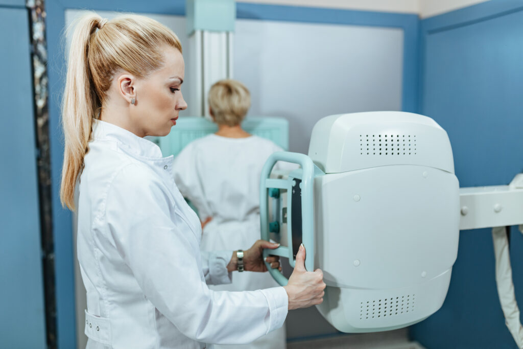
Most major guidelines recommend starting routine mammography at age 40 for women at average risk. However, screening should be adapted to the individual, based on age, risk profile, breast density, and clinical context.
The American College of Radiology recommends annual screening beginning at age 40 for women of average risk and earlier and/or more intensive screening for women at higher-than-average risk. For most women at higher-than-average risk, the supplemental screening method of choice is breast MRI. Women with genetics-based increased risk, those with a calculated lifetime risk of 20% or more, and those exposed to chest radiation at young ages are recommended to undergo Breast MRI surveillance starting at ages 25 to 30 and annual mammography.
Choosing the Appropriate Imaging Method
Different imaging tools play different roles in early detection, and the choice depends on the clinical question being asked.

Looking for expert breast care?
Contact us for consultation and appointment booking
Make an Appointment