Nuclear Medicine

Nuclear Medicine for Diagnostic

Helps detect myocardial ischaemia, hyperthyroidism, urinary tract obstruction and bone abnormalities, while supporting the localisation of abnormal parathyroid glands.

Conventional imaging methods such as X-rays, computed tomography (CT) and magnetic resonance imaging (MRI) show doctors what an organ looks like. Nuclear medicine or scintigraphy scan provides a different perspective: how that organ is functioning.

For a scintigraphy scan, a small amount of a radioactive tracer is introduced into the body and taken up by specific tissues or organs. The resulting images show metabolic activity, blood flow, organ function and cellular activity, helping doctors identify disease processes that may not be visible on CT or MRI alone.

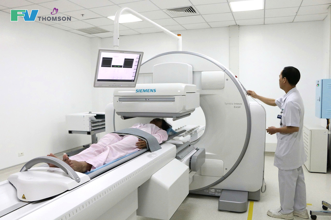
At FV, scintigraphy is performed using a new-generation SPECT gamma camera system. Where appropriate, SPECT can be combined with CT in a single examination (SPECT/CT), allowing doctors to assess both the anatomical location and functional activity of an abnormal area. This combination provides more comprehensive information to support diagnosis and guide appropriate follow-up and treatment.

Frequently Asked Questions About Scintigraphy
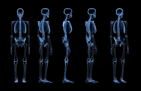
Bone scintigraphy is a whole-body imaging technique that helps doctors identify abnormalities in the skeleton. These may include early bone metastases, bone inflammation, abnormalities following joint replacement, or facial asymmetry.

Bone scintigraphy can detect the spread of cancer to the bones earlier than conventional X-rays in many cases, which may not show a lesion until it reaches a certain size. This earlier detection can be important for people with cancer.

Before your bone scintigraphy scan:

  • You usually do not need to fast and can take your medicines as usual unless your doctor advises otherwise. After the radiotracer is administered, you will need to wait for a period of time for it to accumulate in the bones. During this time, you may be advised to drink extra water and urinate frequently.
  • Tell the nuclear medicine team if you are pregnant, may be pregnant or are breastfeeding.
Thyroid scintigraphy uses a small amount of radioactive material taken up by thyroid cells to produce images that help doctors assess the structure and function of the thyroid gland. Unlike ultrasound, which mainly shows the gland’s structure and thyroid nodules, scintigraphy also provides information about how active different areas of the thyroid are.

Your doctor may recommend thyroid scintigraphy to:

  • Diagnose hyperthyroidism, including distinguishing Graves’ disease from toxic thyroid nodules or thyroiditis
  • Assess thyroid nodules, including hot and cold nodules
  • Assess thyroid uptake before I-131 treatment
  • Identify ectopic thyroid tissue

Thyroid scintigraphy is generally painless. After the scan, you can usually return to your normal activities as advised by your healthcare team.

The nuclear medicine team will provide detailed instructions on how to prepare for your thyroid scan. You should also tell your doctor if:

  • You are taking any medicines, particularly thyroid medicines, vitamins, supplements containing iodine, fish oil, seaweed or kelp products.
  • You have recently had an X-ray or CT scan using iodine-based contrast material, as iodine can affect the thyroid’s uptake of the radiotracer.
  • You are pregnant, may be pregnant or are breastfeeding.
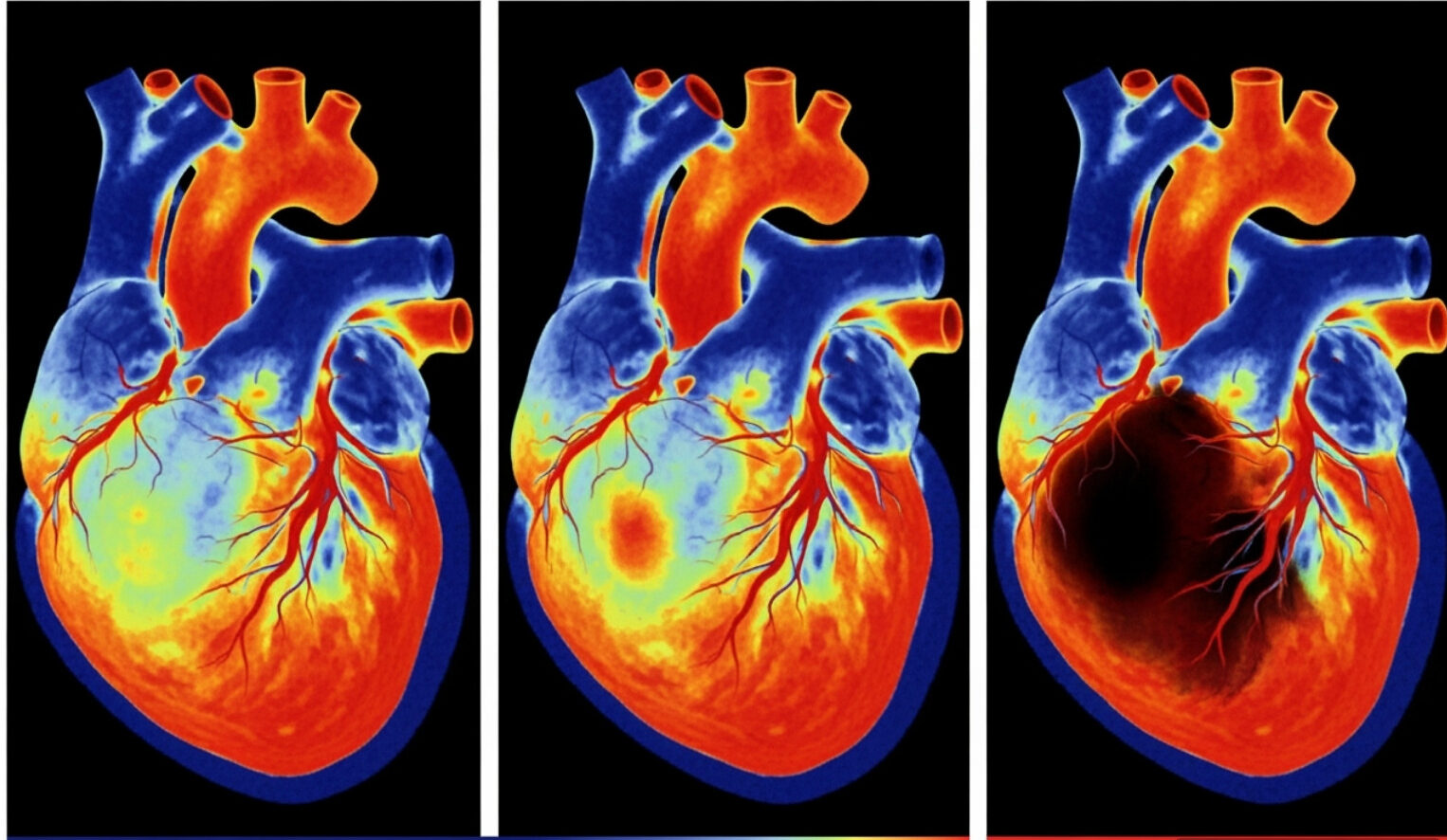
FV is the only hospital in Vietnam performing cardiac amyloidosis scintigraphy, helping detect amyloid deposits in the heart muscle without a biopsy in many cases.

Cardiac amyloidosis occurs when abnormal proteins build up in the heart muscle, gradually impairing its ability to contract and potentially leading to heart failure. The condition can be missed or underdiagnosed because its symptoms may resemble other forms of heart failure. In the past, a definitive diagnosis often required a heart muscle biopsy, which is a specialised and invasive procedure.

Cardiac amyloidosis scintigraphy is a non-invasive test that can help doctors diagnose amyloidosis with a high degree of accuracy, without a biopsy in many cases.

Being able to diagnose amyloidosis without a biopsy is important because many forms of cardiac amyloidosis can now be treated. New therapies targeting the underlying disease process have significantly improved outcomes for patients diagnosed early.

Cardiac amyloidosis scintigraphy may be particularly considered for people with unexplained heart failure, thickened heart walls or other signs suggestive of cardiac amyloidosis.

For cardiac amyloidosis scintigraphy, you will swallow a small amount of a radioactive tracer and wait about one hour for it to be taken up. The scan itself then takes about 15 minutes.

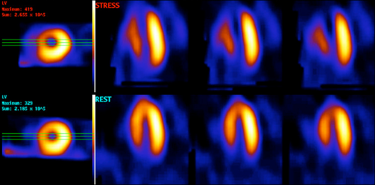
Myocardial perfusion scintigraphy helps doctors assess blood flow to the heart muscle both at rest and during stress. By looking at how the radiotracer is distributed in the heart muscle, doctors can identify areas with adequate blood supply and areas showing reduced perfusion.

This technique is used to assess blood flow through the heart muscle, detect myocardial ischaemia, identify restenosis after stent placement and assess damage from a previous heart attack. It also provides information about heart function, including left ventricular ejection fraction and wall motion, helping doctors determine the most appropriate treatment approach.

Myocardial perfusion scintigraphy is usually performed together with a stress test. Images are taken at two stages – when the heart is at rest and when its activity is increased through exercise or an appropriate alternative. Comparing the two sets of images helps doctors determine whether blood flow to an area of the heart muscle changes when the heart is working harder.

Before your myocardial perfusion scintigraphy:

  • During the 24 hours before the test:
    You may eat normally and drink plain water.
    Do not drink coffee, decaffeinated coffee, tea, herbal tea, decaffeinated tea, soft drinks or any food or drink containing caffeine, including energy drinks.
    Do not consume any food containing chocolate, chocolate flavouring or cocoa.
    Do not use or consume any products containing cannabis (marijuana).
  • Do not eat any food during the four hours before the test.
  • If you have non-insulin-dependent diabetes, please inform the appointment team so that an appropriate appointment time can be arranged.
  • After the stress test, you will be advised to eat within 30–45 minutes. You may bring food from home or have a meal at the hospital canteen.
  • Tell your doctor about all medicines you are taking so that they can advise whether you should continue or temporarily stop any of them.

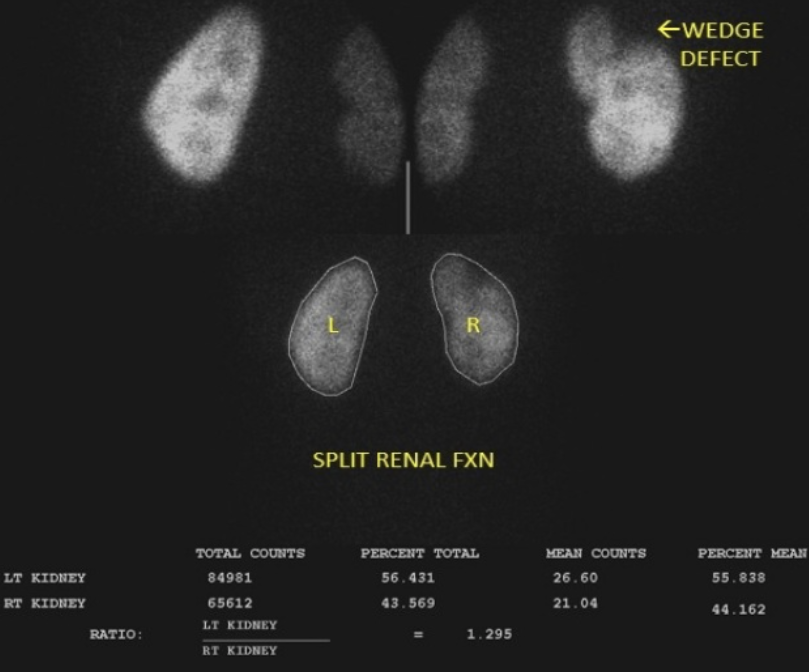
Renal scintigraphy uses a small amount of radioactive material to assess the structure and function of each kidney, as well as how urine is produced and flows through the urinary system. Depending on the purpose of the examination, your doctor may recommend dynamic or static renal scintigraphy to obtain different information about kidney function.

  • Dynamic renal scintigraphy: Assesses the function of each kidney and detects urinary tract obstruction, helping doctors determine whether surgery may be needed. It can also be used to assess kidney function in potential kidney donors before transplantation.
  • Static renal scintigraphy: Assesses renal cortical function and identifies ectopic or malrotated kidneys.

Parathyroid scintigraphy helps localise the parathyroid glands before surgery. This information is important in the treatment of hyperparathyroidism because the glands are small and their anatomical location can vary. A parathyroid gland may sometimes be located in an unusual position in the neck or chest, making it more difficult to locate during surgery.

Parathyroid scintigraphy usually requires no special preparation. You can eat and take your medicines as advised by your doctor. However, before the scan, tell the nuclear medicine team about any medicines, vitamins or supplements you are taking, and let them know if you are pregnant, may be pregnant or are breastfeeding.
You should also bring your PTH and calcium test results, thyroid/parathyroid ultrasound results and any relevant imaging results to help your doctor perform and interpret the scan.

This technique is used in selected cases of breast cancer, melanoma, head and neck tumours and cervical cancer to identify the first lymph node to receive drainage from the tumour site before surgery.

First, a small amount of radiotracer is injected around the tumour. An initial scan is then performed to track the tracer and identify the sentinel lymph nodes.

After the first scan, you will rest in the department for about two hours. A second scan is then performed to confirm the location of the sentinel lymph nodes. Imaging at different time points allows doctors to follow lymphatic drainage and localise the nodes more accurately.

The total procedure time, including the waiting period between the two scans, is about three hours. The actual duration may vary depending on the location of the lesion and the individual’s lymphatic drainage.

Salivary gland scintigraphy assesses the function and secretion of the major salivary glands, including the parotid and submandibular glands.

This technique can be used to assess chronic inflammation or functional impairment of the salivary glands, as well as gland function before and after radiotherapy to the head and neck. Salivary gland scintigraphy can also provide additional information when the cause of persistent dry mouth is unclear, particularly when it is necessary to assess whether reduced salivary secretion is contributing to the symptoms.

Testicular scintigraphy helps doctors assess blood flow to the testicles and distinguish orchitis (infection or inflammation) from testicular torsion, a surgical emergency requiring immediate treatment, in patients with acute scrotal pain when clinical examination alone is inconclusive.

In people with differentiated thyroid cancer who have undergone thyroidectomy, an I-131 whole-body scan is used to assess for residual thyroid tissue or metastases. The results can help doctors determine whether low- or high-dose I-131 treatment is appropriate and assess the response to treatment.

A whole-body scan is usually performed about 4–6 weeks after surgery, as advised by your doctor.

To help ensure accurate scan results, you will need to follow certain preparation instructions:

  • Stop taking thyroid hormone about four weeks before the scan, as directed by your doctor.
  • Limit foods high in iodine and seafood for about two weeks before the scan.

Has your doctor recommended a scintigraphy scan?

Contact us to schedule your scan or ask how to prepare.

Make an Appointment
MessengerZalo