



Suffering from frequent chest pain, high blood pressure, and fainting, Ms. P.T.L., 52 years old, from An Giang, was diagnosed with coronary anaemia. However, after decades of treatment, her condition remained unresolved.
Recently, doctors at FV Hospital accurately diagnosed her with congenital aortic coarctation and successfully treated her through a stent placement procedure.
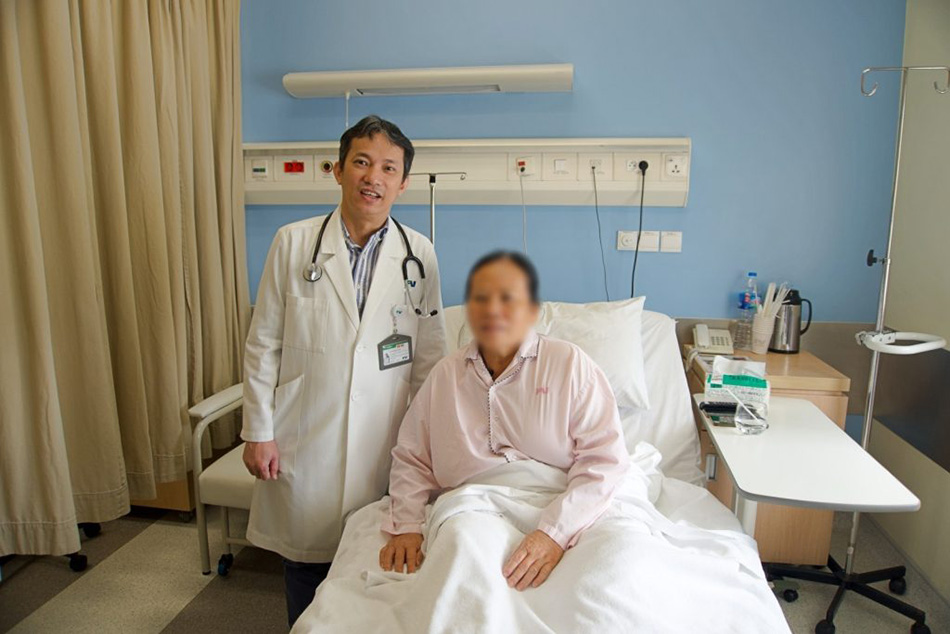
Thanks to Dr Ho Minh Tuan, PhD, who identified and directly treated Ms P.T.L. with a stent placement, she has finally been freed from the aortic coarctation that had afflicted her for over 50 years
Initially Diagnosed with Coronary Anaemia, Later Revealed as Aortic Coarctation
Since childhood, Ms P.T.L. had frequently experienced sudden chest pain. After consulting multiple doctors, she was diagnosed with coronary anaemia. Despite being prescribed medication, her condition persistently relapsed, leading to shortness of breath, chest pain, high blood pressure, and frequent fainting spells that required emergency care.
In November 2024, as her condition deteriorated, her family brought her to Ho Chi Minh City for emergency treatment. Following a referral, she sought care at FV Hospital, consulting with Ho Minh Tuan, MD, PhD, Head of the Cardiology and Interventional Cardiology Department, in the hope of finally finding a lasting solution to the illness she had endured for over 50 years.
Dr Tuan observed that the patient had extremely high blood pressure, with spikes reaching 200 mmHg, along with symptoms of heart failure causing fatigue and leg weakness, which made movement challenging. “Typically, patients with high blood pressure, especially those with fluctuating hypertension and symptoms of heart failure and leg weakness, are suspected to have congenital aortic coarctation. After conducting several tests, particularly ultrasound and CT angiography, the results confirmed that the patient had congenital aortic coarctation,” shared Dr Tuan.
Dr Tuan explained that congenital aortic coarctation leads to episodic high blood pressure, significantly increasing the risk of stroke caused by cerebral haemorrhage. The condition also leads to impaired blood flow, forcing the heart to work harder and potentially leading to heart failure over time. Furthermore, reduced blood flow to the lower legs causes fatigue during movement, as the legs do not receive an adequate blood supply.
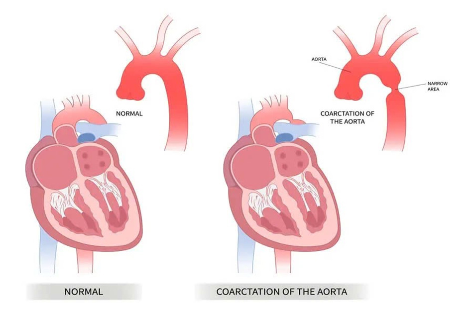
Illustration of congenital aortic coarctation causing restricted blood flow from the heart to the organs
The aortic coarctation that Ms L. suffers from is a congenital heart condition that occurs in approximately 2 out of every 10,000 newborns. From birth, she had a narrowing in the segment of the aorta, the main artery responsible for carrying blood from the heart to the rest of the body. Due to its critical location, blood pressure before the narrowing is abnormally high, directly affecting blood flow to the brain and increasing the risk of cerebral haemorrhage – a condition Ms L. has been fortunate to avoid. Beyond the narrowing, blood pressure drops significantly, resulting in insufficient blood supply to the limbs and lower body.
Aortic Stenting to Treat Coarctation of the Aorta
To treat aortic coarctation, stent placement is the preferred method due to its proven safety and effectiveness. However, in Ms L.’s case, the procedure was more complex as the stent needed to be precisely placed at the coarctation site. Typically, the access point for stent placement has a diameter of 2-3 mm. In this instance, Dr Tuan had to enlarge the access diameter to 5-6 mm using a specialised closure device, preventing uncontrolled bleeding.
Since the stent is placed in the aorta, its larger size necessitates a wider access point in the thigh. “This is a special type of stent: regular stents have one balloon that expands when it reaches the desired location, pressing the stent against the vessel wall. This particular stent, however, has two balloons: the small balloon is inflated first, followed by the larger balloon, which ensures the stent fits properly against the vessel wall,” Dr Tuan explained.
This procedure requires careful handling to avoid vascular trauma, which could result in a tear and uncontrolled bleeding. Furthermore, the stent must be placed exactly as planned, any misalignment could cause it to shift out of position, compromising its effectiveness.
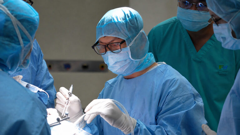
The stent placement for Ms L. on November 26th proceeded smoothly. Dr Ho Minh Tuan performed the balloon dilation and stent placement with the assistance of a Digital Subtraction Angiography (DSA) system. The procedure was successfully completed in just 60 minutes, and the patient remained fully awake throughout.
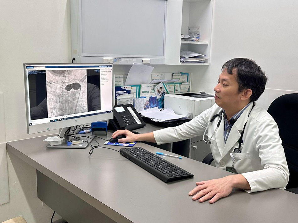
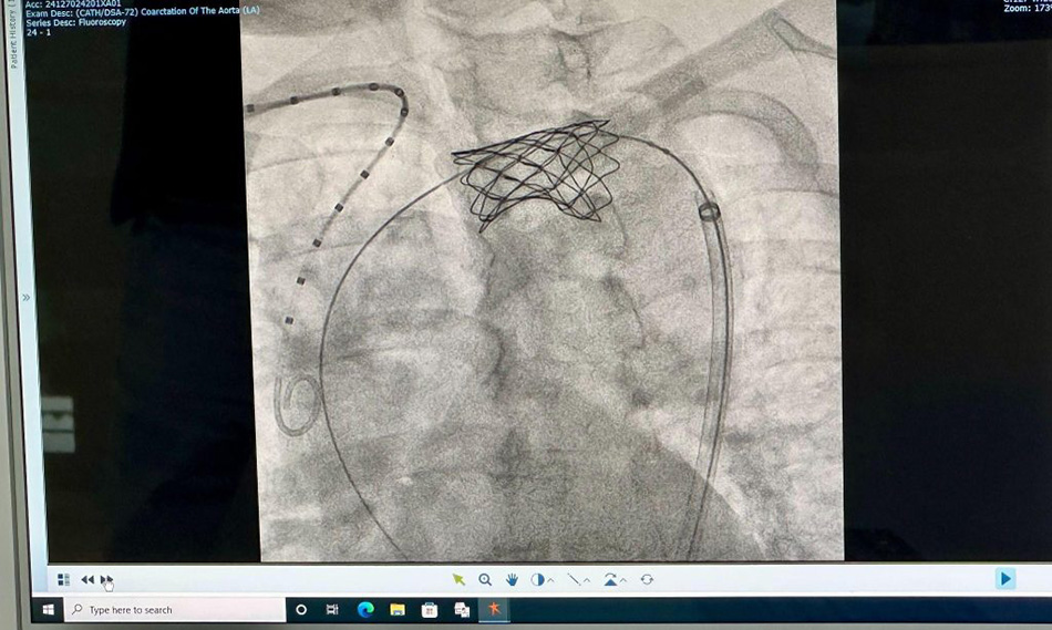
The image of the aortic stent captured after successful placement
“Dr Tuan from FV provides excellent treatment!”
“Before the procedure, I was really anxious, but Dr Tuan reassured and comforted me a lot. During the procedure, he was very gentle and careful, and I was fully awake, so I could feel all the actions he was taking. After the procedure, I felt fine, with no discomfort at all. Dr Tuan treated me excellently! Thanks to his diagnosis and treatment, I now have the chance to live a long life,” Ms P.T.L. shared excitedly.
After the procedure, the patient’s blood pressure levels stabilised, and the symptoms of leg pain during movement disappeared. Blood circulation improved significantly, and the patient was discharged two days later.
Ms L. shared that throughout her treatment and follow-up at FV Hospital, she received excellent care, including meals, hygiene, and bathing. “The international hospital environment is very clean, and I feel like it was truly worth the money,” Ms L. expressed with gratitude before being discharged.
To learn more about the treatment of aorta coarctation and other cardiovascular diseases, please contact the Cardiology and Interventional Cardiology Department at FV Hospital at: (028) 54 11 33 33.
Related News
See moreRelated News
Abdominal pain misdiagnosed as digestive issue: 23-year-old woman diagnosed with ovarian torsion
While studying abroad in South Korea, N.T.T.M. (23) experienced unusual pain in her lower right abdomen. She sought treatment at a local hospital and received medication for a suspected digestive issue. After returning to Vietnam to visit her family, the pain recurred and worsened. On her friends’ advice, M. visited FV Hospital for evaluation. She […]
Advanced robotic surgery at FV Hospital removes tumour near aorta
Surgeons at FV Hospital used the da Vinci Xi robotic surgical system to successfully remove a fibrous tumour located in a difficult-to-access area next to the aorta in a 46-year-old woman. Ms Nguyen Thuy Phuong Dung recovered well after surgery to remove the fibrous tumour from her left chest. Image: FV When a subtle chest […]
Boy with ‘boneless arm’ and decade-long journey to find hope
A jump at the age of six left young D.V.C. with devastating consequences: a left forearm almost entirely devoid of skeletal architecture, facing the prospect of limb amputation through joint disarticulation. For more than ten years, hope faded as every attempt at recovery stalled. It was only through the intervention of the Children of Vietnam […]
Travelling halfway across the world for specialist spinal stenosis treatment
A Canadian woman who had nearly lost the ability to walk due to spinal canal stenosis was successfully treated at FV Hospital and was able to return home just one week after surgery. More than thirty years ago, Ms M.C. (49, Canada) underwent scoliosis correction surgery. In the past year, she developed severe back pain […]
Abdominal pain misdiagnosed as digestive issue: 23-year-old woman diagnosed with ovarian torsion
Advanced robotic surgery at FV Hospital removes tumour near aorta
Boy with ‘boneless arm’ and decade-long journey to find hope

