




For the first time in Southern Vietnam, a complex liver tumour has been successfully treated using a combination of biopsy and radiofrequency ablation (RFA) guided by a next-generation Fusion and Navigation Ultrasound Imaging system at FV Hospital. The minimally invasive procedure was performed under local anaesthesia, completed quickly, and allowed the patient to recover and be discharged after just one day of observation.
Fusion and ultrasound imaging enables precise localisation of liver tumours
Fusion and Navigation Imaging is an advanced ultrasound-guided technology that enables physicians to accurately identify liver lesions located in difficult-to-access areas. It facilitates precise biopsy and radiofrequency ablation of liver tumours without the need for open surgery. As a result, the procedure is faster, safer, and allows patients to recover more quickly, with most being discharged after only one to two days of monitoring.
Ms T.T.X.H. (51, from Tay Ninh Province) was found to have a liver tumour while undergoing chemotherapy for upper abdominal cancer. The tumour was located in an extremely challenging position and could not be visualised using conventional ultrasound equipment. According to her doctors, without timely treatment, the tumour could continue to grow, spread locally, and metastasise to surrounding organs, significantly impairing liver function and reducing her quality of life.
In similar cases, surgical liver resection would normally be recommended. However, this invasive approach requires general anaesthesia, causes greater postoperative discomfort, and involves a longer recovery period. To provide a less invasive alternative, FV’s interventional team utilised the Philips PercuNav Fusion and Navigation System, integrated into the hospital’s high-end ultrasound platform.
Simply put, the technology functions like a dedicated GPS navigation system for physicians. It automatically overlays detailed CT or MRI images onto live ultrasound during the procedure, creating a real-time roadmap for accurate intervention. One of its key advantages is its automatic image registration capability. Instead of relying on time-consuming manual alignment, the AI-powered system automatically recognises the patient’s liver anatomy and vascular structures in less than one minute, generating a highly accurate navigation map that guides the physician throughout the procedure.
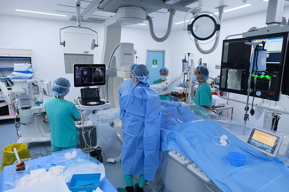
Specialists perform Fusion and Navigation Imaging in FV Hospital’s state-of-the-art cathlab
This breakthrough technology delivers significant clinical benefits. It reduces the number of needle repositioning attempts by up to 64%, minimising the risk of complications for patients. It also decreases the need for repeat CT scans during the procedure by 45%, significantly reducing radiation exposure while shortening procedure time.
“In addition, this minimally invasive approach offers several advantages. Patients remain fully awake throughout the procedure and experience little to no pain or discomfort at the treatment site afterwards,” said FV’s interventional radiology team.
Where advanced technology meets patient trust
The liver tumour ablation procedure lasted just over two hours and was performed in FV’s state-of-the-art interventional suite under local anaesthesia, eliminating the need for general anaesthesia. Guided by the real-time navigation system, the medical team accurately inserted the needle through the skin into the centre of the tumour before delivering radiofrequency energy of up to 100°C to completely and safely destroy the liver lesion.
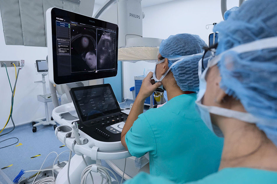
Specialists reviewing the Fusion and Navigation Imaging system
It is natural for patients to feel anxious before undergoing an interventional procedure, especially when the lesion is hidden in a difficult-to-reach location. However, thorough counselling from the medical team, combined with their expertise and the precision of the real-time navigation technology, gave Ms X.H. the confidence to proceed with treatment. “I wasn’t sure whether there were any treatment options left because my case was quite complicated. But after the procedure, I can truly say it was a success,” she said.
Thanks to the technology’s ability to minimise damage to healthy liver tissue and support faster recovery, Ms X.H. was able to resume normal daily activities and was discharged after just one day of close observation. The dedicated care and continuous support from FV’s medical team also helped ease her concerns and made her feel reassured throughout the treatment process.
Reflecting on her experience, Ms X.H. said she felt fortunate to have benefited from this advanced technology and the attentive care provided at FV Hospital. She hopes her successful treatment will pave the way for more patients to benefit from the same innovative approach in the future.
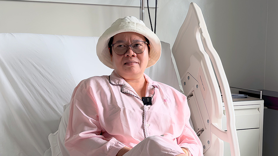
Ms T.T.X.H. made a rapid recovery following the procedure
The success of the first Fusion and Navigation Imaging-guided liver intervention in Southern Vietnam not only delivered an excellent outcome for the patient, but also reinforced FV Hospital’s position as a pioneer in adopting advanced medical technologies. It marks an important milestone that will support the expansion of this technology into a wider range of complex interventional procedures, further enhancing the quality of patient care.
| Fusion and Navigation Imaging is currently being used at FV Hospital for complex liver interventions, particularly for liver lesions and tumours located in difficult-to-access areas. According to FV’s specialists, the technology also holds strong potential for broader clinical applications in the future, including image-guided percutaneous procedures for breast and prostate tumours, as well as targeted (focal) biopsies to accurately sample suspicious areas where cancer recurrence is suspected. Its versatility offers more patients access to advanced, minimally invasive treatment options. Book an appointment today to consult with the specialists at the Imaging & Interventional Radiology Department, FV Hospital. Address: 6 Nguyen Luong Bang Street, Tan My Ward (formerly District 7), Ho Chi Minh City, Vietnam Hotline: (+84) 28 3511 3333 |
Related News
See moreRelated News
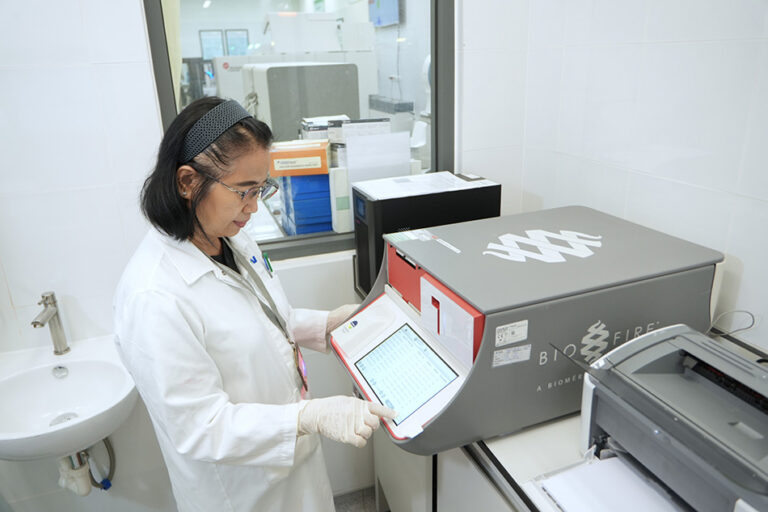
Quickly identify pathogens with 4 molecular diagnostic systems at FV
With four advanced rapid molecular diagnostic systems, BioFire® FilmArray®, QIAGEN QIAstat-Dx®, Cepheid GeneXpert®, and Sansure iPonatic III, the FV Hospital Laboratory & Blood Bank Department offers a wide range of rapid testing to help identify the bacteria, viruses and other infectious pathogens, helping your doctor make timely and informed treatment decisions. FV Hospital Laboratory & […]
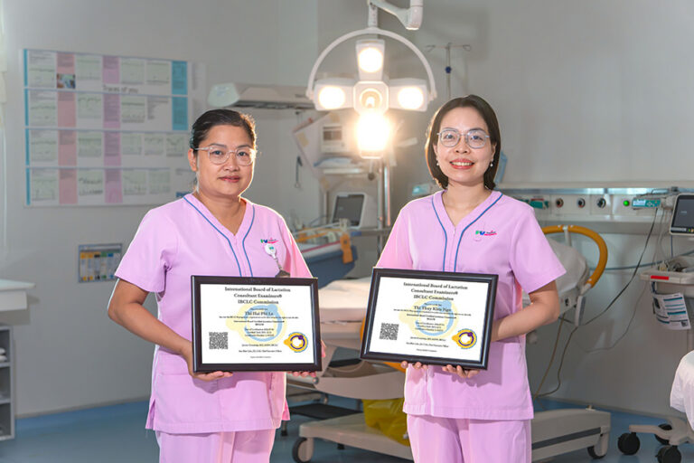
Two Vietnamese midwives attain international IBCLC certification in breastfeeding counselling
Two midwives from FV Thomson’s Maternity Department have earned IBCLC (International Board Certified Lactation Consultant) certification, the international credential for breastfeeding support specialists. This achievement represents a significant clinical milestone in infant feeding support at FV Hospital, driven by the real-world complexities these practitioners encounter daily when supporting mothers and their newborns. Beyond certification: A […]

Nutcracker Syndrome: How a one-hour stent placement ended a year of abdominal pain
FV Hospital successfully treated a complex case of nutcracker syndrome using minimally invasive endovascular stenting, sparing the patient major open surgery. Persistent abdominal pain linked to renal vein entrapment For over a year, 41-year-old Ms V.T.K.P. from Ho Chi Minh City experienced persistent, dull lower abdominal pain that worsened with prolonged sitting or standing. As […]
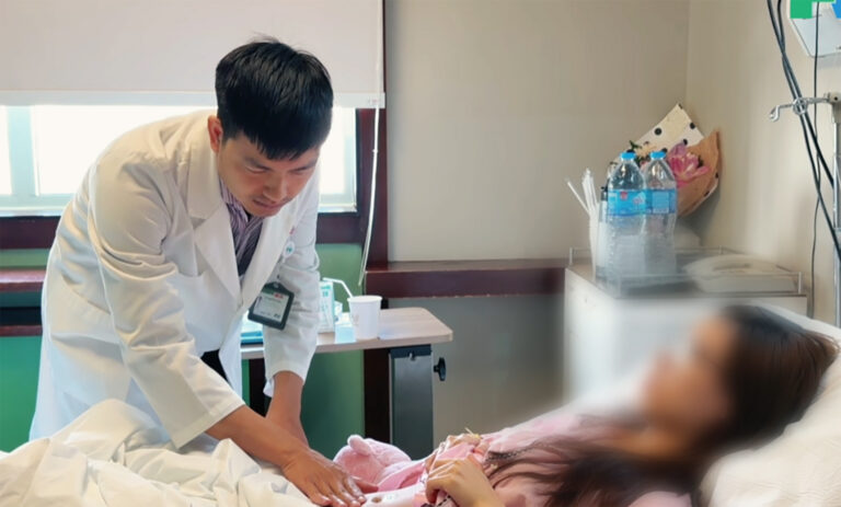
Abdominal pain misdiagnosed as digestive issue: 23-year-old woman diagnosed with ovarian torsion
While studying abroad in South Korea, N.T.T.M. (23) experienced unusual pain in her lower right abdomen. She sought treatment at a local hospital and received medication for a suspected digestive issue. After returning to Vietnam to visit her family, the pain recurred and worsened. On her friends’ advice, M. visited FV Hospital for evaluation. She […]
Quickly identify pathogens with 4 molecular diagnostic systems at FV
Two Vietnamese midwives attain international IBCLC certification in breastfeeding counselling
Nutcracker Syndrome: How a one-hour stent placement ended a year of abdominal pain

