




After nearly 10 days of treatment at two healthcare facilities without a clear diagnosis for her recurrent pneumothorax, a 23-year-old female patient was successfully treated by doctors at FV Hospital using video-assisted thoracoscopic surgery.
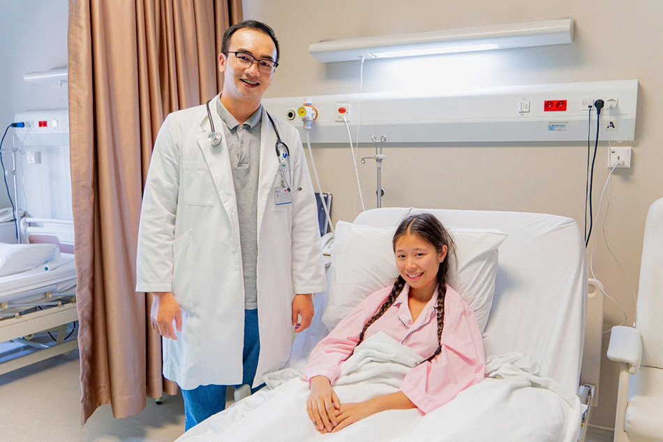
Dr Pham Nguyen Thanh Nam with the patient following her treatment at FV Hospital’s Vascular and Endovascular Surgery Department.
M.J., 23 years old French national of of Vietnamese origin, working in Vietnam’s tourism sector, was admitted to FV Hospital with sudden right-sided chest pain and shortness of breath. She was diagnosed with spontaneous pneumothorax and underwent chest tube drainage at another healthcare facility. However, after several days of observation, her chest pain persisted despite chest tube drainage, while the underlying cause remained unclear.
Anxious about her prolonged illness and facing communication barriers due to her limited fluency in Vietnamese, M.J. turned to FV Hospital.
Rather than focusing solely on diagnostic test results, FV doctors reviewed her medical history in detail, from the onset and progression of her symptoms to relevant aspects of her daily life. A crucial detail emerged: her episodes of chest pain coincided with her menstrual cycle.
Following a multidisciplinary consultation, Dr Luong Ngoc Trung, MSc, Specialist Level II, Thoracic and Vascular Surgeon and Head of Vascular and Endovascular Surgery Department, confirmed that the patient had catamenial pneumothorax, a form of spontaneous pneumothorax associated with the menstrual cycle. This is a form of spontaneous pneumothorax associated with menstruation in women of reproductive age, typically occurring at the onset of menstruation or within 72 hours thereafter.. Studies indicate that it accounts for only about 3–6% of spontaneous pneumothorax cases in women, though this rate can be higher in specialised treatment cohorts, making it a rare condition that can easily be overlooked.
According to Dr Trung, the condition is usually linked to thoracic endometriosis, where endometrial tissue grows on the diaphragm or pleural lining. During each menstrual cycle, these lesions may contribute to air entering the pleural space from the abdominal cavity into the pleural space, causing the lung to partially or completely collapse. Because its symptoms can closely resemble those of spontaneous pneumothorax, many cases are overlooked, increasing the risk of recurrent episodes associated with the menstrual cycle.
For M.J., definitive treatment was particularly important because her job requires frequent air travel. A recurrent pneumothorax during a flight could pose a serious health risk.
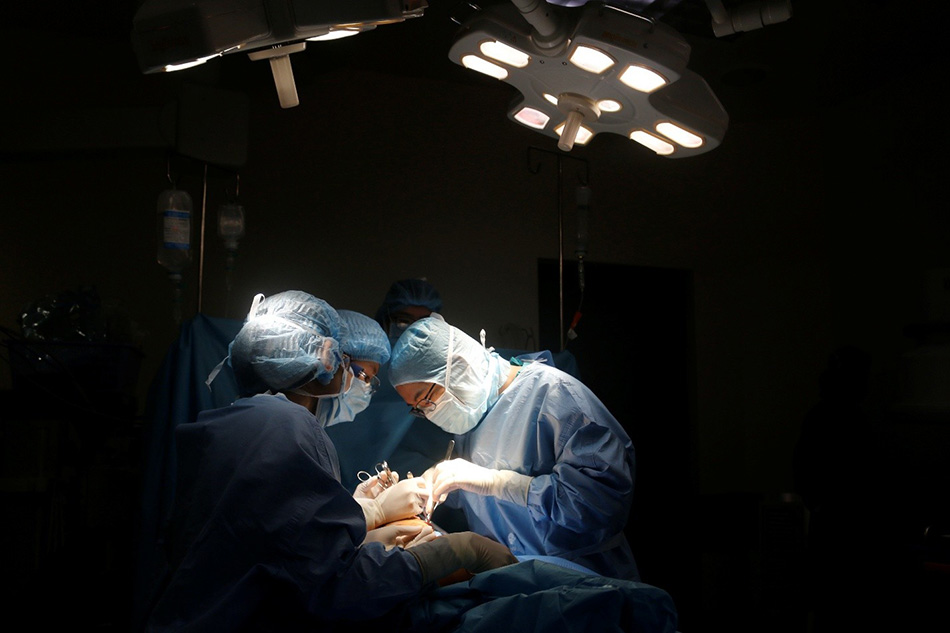
The surgical team performing Video-Assisted Thoracoscopic Surgery (VATS).
Following a comprehensive assessment, the surgical team opted for Video-Assisted Thoracoscopic Surgery (VATS). Using an endoscopic camera, the surgeons examined the pleural cavity, treated the suspected lesions, and performed pleurodesis to minimise the risk of recurrence. Findings during the operation showed no ruptured air sacs (pulmonary bullae) or other lung lesions responsible for air leaks.
VATS is widely used to treat recurrent pneumothorax, particularly when thoracic endometriosis is suspected.
Just a few hours after surgery, M.J. was able to sit up and walk around with ease. Two days later, she was discharged in stable condition. “I knew I was finally being treated for the right condition,” she shared. “The relief was completely different from the uncertainty and anxiety I felt during the previous ten days.”
According to Dr Luong Ngoc Trung, MSc, Specialist Level II, despite its rarity, catamenial pneumothorax should be considered in young women with recurrent pneumothorax, especially when symptoms occur within approximately 72 hours before or after menstruation. Taking a detailed patient history is as vital as diagnostic imaging in helping doctors identify the correct cause and choose the appropriate treatment strategy. The condition has also been published in a prestigious international medical journal by a group of doctors from FV Hospital. The journal, Radiology Case Reports, is published by Elsevier.
https://doi.org/10.1016/j.radcr.2025.04.033
| FV Hospital is one of the centre in Vietnam with a high rate of VATS utilisation, with the technique used in more than 95% of thoracic surgery cases. Single-port VATS (Uniport VATS) is routinely used for many procedures, offering a minimally invasive approach with less postoperative pain and faster recovery. For complex cases, FV also offers robotic-assisted thoracic surgery using the da Vinci Xi system, helping improve surgical precision and optimise treatment outcomes in complex cases.. |
Related News
See moreRelated News

Nutcracker Syndrome: How a one-hour stent placement ended a year of abdominal pain
FV Hospital successfully treated a complex case of nutcracker syndrome using minimally invasive endovascular stenting, sparing the patient major open surgery. Persistent abdominal pain linked to renal vein entrapment For over a year, 41-year-old Ms V.T.K.P. from Ho Chi Minh City experienced persistent, dull lower abdominal pain that worsened with prolonged sitting or standing. As […]
Abdominal pain misdiagnosed as digestive issue: 23-year-old woman diagnosed with ovarian torsion
While studying abroad in South Korea, N.T.T.M. (23) experienced unusual pain in her lower right abdomen. She sought treatment at a local hospital and received medication for a suspected digestive issue. After returning to Vietnam to visit her family, the pain recurred and worsened. On her friends’ advice, M. visited FV Hospital for evaluation. She […]
Advanced robotic surgery at FV Hospital removes tumour near aorta
Surgeons at FV Hospital used the da Vinci Xi robotic surgical system to successfully remove a fibrous tumour located in a difficult-to-access area next to the aorta in a 46-year-old woman. Ms Nguyen Thuy Phuong Dung recovered well after surgery to remove the fibrous tumour from her left chest. Image: FV When a subtle chest […]
Boy with ‘boneless arm’ and decade-long journey to find hope
A jump at the age of six left young D.V.C. with devastating consequences: a left forearm almost entirely devoid of skeletal architecture, facing the prospect of limb amputation through joint disarticulation. For more than ten years, hope faded as every attempt at recovery stalled. It was only through the intervention of the Children of Vietnam […]
Nutcracker Syndrome: How a one-hour stent placement ended a year of abdominal pain
Abdominal pain misdiagnosed as digestive issue: 23-year-old woman diagnosed with ovarian torsion
Advanced robotic surgery at FV Hospital removes tumour near aorta

