




FV Hospital recently treated a challenging case of severe headache and drooping eyelid caused by chronic hypertrophic pachymeningitis. This condition makes the dura mater inflamed and thickened, which can appear as a brain tumour on scans.
At first, Mr D.N.G. (24, from Vinh Long Province) presented with occasional headaches and fatigue. MRI scans revealed a small lesion at the skull base and cavernous sinus, a region commonly associated with meningiomas. Because the lesion was still small and thin, he received medication and was advised to undergo regular monitoring.
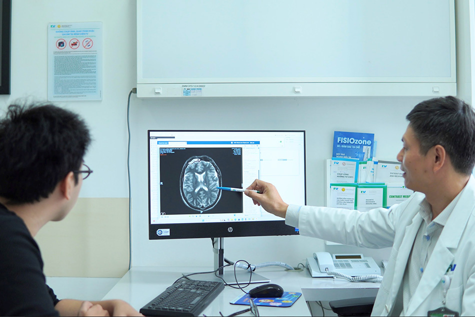
Dr Tran Luong Anh, Specialist Level II, discussing brain lesion imaging with the patient. Image: FV Hospital
A month later, his headaches intensified, and he developed left eyelid drooping and double vision. Mr G. sought care at FV Hospital, where Dr Tran Luong Anh, Specialist Level II, Head of Neurosurgery and Spine Surgery, said: “There were features suggesting this lesion did not fully fit the diagnosis of a meningioma.”
Suspecting an intracranial inflammatory process, the clinical team ordered comprehensive blood tests, a lumbar puncture for cerebrospinal fluid analysis, and other investigations. However, none of the investigation results revealed any significant abnormalities.
Surgical exploration uncovers a ‘pseudotumour’ adhering to the cavernous sinus
With no clear cause identified through non-invasive testing, Dr Luong Anh proceeded with surgical decompression of the affected cranial nerve to relieve symptoms and obtain tissue for histopathological diagnosis.
During the craniotomy, Dr Luong Anh and his team used an operating microscope to observe that the dura mater was inflamed. They found extensive dural inflammation, with reddish-pink tissue firmly attached to the dura mater, forming a distinct “pseudotumour” that resembles a true tumour in appearance.
“This inflammatory tissue had directly invaded the wall of the cavernous sinus and compressed the oculomotor nerve, resulting in the patient’s ptosis and diplopia,” Dr Tran Luong Anh, Specialist Level II, explained.
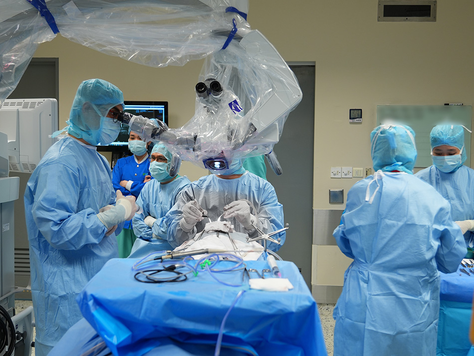
Dr Tran Luong Anh, Specialist Level II, and the surgical team performing a craniotomy. Image: FV Hospital
Histopathological analysis confirmed that Mr G. had a rare autoimmune inflammatory condition, where immune system dysfunction leads to chronic inflammation. In some cases, this persistent response causes significant tissue thickening, resulting in a “pseudotumour” that closely resembles a true tumour on imaging.
After confirming the autoimmune cause, Mr G. began appropriate medication. Within two weeks, his left eye reopened and his ptosis nearly resolved.
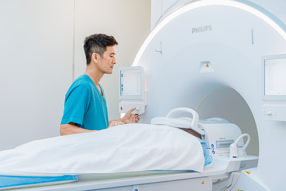
The patient undergoes a comprehensive clinical and radiological evaluation before treatment decisions are made. Image: FV Hospital
Dr Tran Luong Anh emphasised that not every lesion that appears tumour-like on MRI represents a true intracranial neoplasm.
“In complex clinical presentations, physicians must synthesise clinical signs, advanced neuroimaging, laboratory diagnostics, and, when indicated, tissue histopathology to determine the true nature of the lesion. Achieving an accurate diagnosis ensures the patient receives targeted, aetiology-based treatment, avoids unnecessary radical surgical resections, and significantly improves long-term clinical prognosis,” Dr Luong Anh emphasised.
At FV Hospital, complex neurological cases are managed through a multidisciplinary approach involving Neurosurgery, Neurology, Diagnostic Imaging, and Pathology. This collaboration enables specialists to thoroughly assess unusual cases, especially rare tumour-mimicking lesions, and provide each patient with a tailored and effective treatment plan.
To learn more about neurological care and surgical services at FV Hospital, please visit 6 Nguyen Luong Bang Street, Tan My Ward (formerly District 7), Ho Chi Minh City, or call (028) 3511 3333. Emergency Hotline: (028) 3511 3500.
Related News
See moreRelated News

Nutcracker Syndrome: How a one-hour stent placement ended a year of abdominal pain
FV Hospital successfully treated a complex case of nutcracker syndrome using minimally invasive endovascular stenting, sparing the patient major open surgery. Persistent abdominal pain linked to renal vein entrapment For over a year, 41-year-old Ms V.T.K.P. from Ho Chi Minh City experienced persistent, dull lower abdominal pain that worsened with prolonged sitting or standing. As […]
Abdominal pain misdiagnosed as digestive issue: 23-year-old woman diagnosed with ovarian torsion
While studying abroad in South Korea, N.T.T.M. (23) experienced unusual pain in her lower right abdomen. She sought treatment at a local hospital and received medication for a suspected digestive issue. After returning to Vietnam to visit her family, the pain recurred and worsened. On her friends’ advice, M. visited FV Hospital for evaluation. She […]
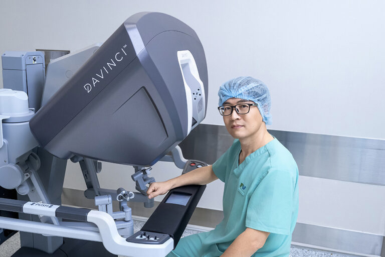
Advanced robotic surgery at FV Hospital removes tumour near aorta
Surgeons at FV Hospital used the da Vinci Xi robotic surgical system to successfully remove a fibrous tumour located in a difficult-to-access area next to the aorta in a 46-year-old woman. Ms Nguyen Thuy Phuong Dung recovered well after surgery to remove the fibrous tumour from her left chest. Image: FV When a subtle chest […]
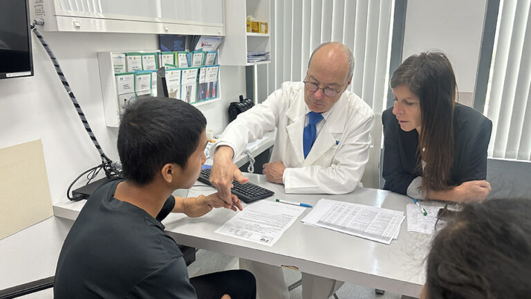
Boy with ‘boneless arm’ and decade-long journey to find hope
A jump at the age of six left young D.V.C. with devastating consequences: a left forearm almost entirely devoid of skeletal architecture, facing the prospect of limb amputation through joint disarticulation. For more than ten years, hope faded as every attempt at recovery stalled. It was only through the intervention of the Children of Vietnam […]
Nutcracker Syndrome: How a one-hour stent placement ended a year of abdominal pain
Abdominal pain misdiagnosed as digestive issue: 23-year-old woman diagnosed with ovarian torsion
Advanced robotic surgery at FV Hospital removes tumour near aorta

