




Liver primary cancer is common in Vietnam. It often develops in patients with hepatitis: the hepatitis virus triggers cirrhosis and then hepatocellular carcinoma (HCC). Open surgery to remove tumours often causes heavy blood loss and large sutures, which in turn may lead to high risk of infection and complications, some of which may even be life threatening. Currently, the most renowned hospitals in the world are utilising less invasive surgical methods to treat liver cancer. FV Hospital’s General Surgery Department provided successful surgical treatments to many complicated cases with multiple and/or large liver tumours.
Ultrasonic aspirator
French patient, Mr. J.B, 45, had colon cancer with hepatic metastatic tumours. In this case, surgery was the only option to save his life. Dr Do Minh Hung, Head of General Surgery, FV Hospital, decided to operate on Mr. J to remove both the colon cancer and hepatic metastatic tumours via minimally invasive coelioscopy procedure (introducing an endoscope through a small incision in the abdomen).
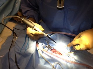
As the tumours were fairly large, an ultrasonic aspirator was used doing the surgery to remove these tumours and to improve surgical safety and efficiency. After six hours in OT, all of the metastatic tumours and colon tumours had been removed. The patient experienced no post-surgical complications.
The ultrasonic aspirator is a versatile system that allows exceptional control of soft tissue and fine dissection to remove tumours present in the liver. Utilising this surgical device in liver cancer treatment has many benefits for patients, including greater accuracy in the removal of tumours without touching the surrounding healthy tissues or harming the tiniest blood vessels, minimising the risk of bleeding and complications after surgery, thereby aiding a shorter recovery time.
Dr Minh Hung said: “The big advantage of using an ultrasonic aspirator in surgical treatments for liver cancer is that it increases safety for patients who require surgical treatment for liver cancer. This special surgical device helps to comprehensively remove cancer and minimise damage to the patient’s liver, as well as foster liver function restoration at the same time.”
FV Hospital has invested in the installation of a Sonopet ultrasonic aspirator and the device has already been used in several successful liver cancer surgeries, both to treat primary malignancy and metastatic cancers.
Chemoembolisation
If surgical treatment fails to remove a tumour completely, utilization of chemoembolisation can save the life of a patient. This effective liver cancer treatment procedure is performed at FV Hospital by Head of Imaging Dr Pierre Jaillot.
Chemoembolisation of the liver involves the injection of chemotherapy agents directly into the arteries to block or restrict the blood supply feeding the liver tumour.
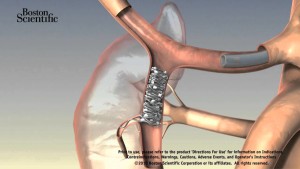
The liver has two blood supplies – the portal vein that feeds most normal liver cells and the hepatic artery that feeds cancer cells in the liver. This unique feature of the liver allows doctors to block the branches of arteries feeding the tumours and kill the cancer cells while sparing the healthy tissues as they continue to receive nutrients from the portal vein.
Chemoembolisation is performed by placing a small catheter into the artery that supplies blood to the liver via the groin. The chemotherapeutic drugs (anti-cancer drug) are then delivered through the catheter along with a blood vessel occluding agent to the blood vessel feeding the tumour. The result is that a highly concentrated dose of anti-cancer drug is delivered to the tumour site to kill the cancerous cells. At the same time, tumours are starved of blood supply as blood vessels are partially blocked with the occluding agent.
This procedure is recommended for cases in which surgery is impossible to remove the tumour located inside liver, increasing the survival rate and living quality for patients. This is one of the most advanced treatments for liver cancer being successfully applied worldwide, including at FV Hospital.
The duration of this procedure is typically from 45 minutes to two hours. Patients remain awake during the procedure. Within a few days after the procedure, patients can leave the hospital. If treatment is successful, a significant decrease in tumour size will be seen three months following the procedure. In some cases, tumours may disappear completely.
The advantages of using chemoembolisation to treat liver cancer is that there is no incision, less pain and a faster recovery following the procedure; patients can also undergo this procedure many times.
Liver cancer is a critical disease and is one of the leading causes of death in Vietnam and around the world. With advances in contemporary medicine, liver cancer patients still have a chance to be cured and live a normal life.
Related Articles
Related Articles

Virtual Colonoscopy (CT Colonography)
WHAT IS A VIRTUAL COLONOSCOPY? Virtual colonoscopy, also called CT colonography, is a method of screening the colon for precancerous polyps using a CT scanner. CT scanning is a non-invasive medical test that combines special x-ray equipment with sophisticated computers to produce multiple images of the inside of the body. CT colonography provides an interior […]
Colonoscopy
COLONOSCOPY OVERVIEW Colonoscopy is an investigation that allows your doctor to look at the inner lining of your large intestine (rectum and colon) using a thin, flexible tube called a colonoscope. A colonoscopy helps find ulcers, polyps, tumors, and areas of inflammation or bleeding. During a colonoscopy, tissue samples can be collected (biopsy) and abnormal […]
FIBROSCAN
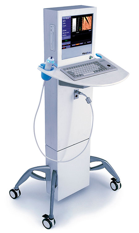
FibroScan FibroScan is the first and only non-invasive device for diagnosis and monitoring of hepatic fibrosis. Why do we need to quantify liver fibrosis? In its initial stages, liver fibrosis develops silently and often causes no symptoms. Early diagnosis and treatment will help prevent the disease from progressing to liver cancer, liver function failure, or […]
Endoscopic retrograde cholangiopancreatography (ERCP)
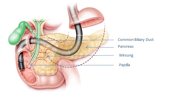
What is ERCP? Endoscopic retrograde cholangiopancreatography, or ERCP, is an endoscopic technique that is used to examine and treat problems in the bile and pancreatic ducts, and the gallbladder. The bile ducts are channels that drain bile from the liver and gallbladder; the pancreatic ducts drain the pancreas. Both types of ducts empty into the […]
Virtual Colonoscopy (CT Colonography)
FIBROSCAN

