




Mr Nguyen Van Hai, 66, was diagnosed with a lung tumour measuring approximately 17 mm. However, surgery was particularly complex because of his 40-year smoking history and a history of pulmonary tuberculosis around 30 years ago. Scarring caused by the previous tuberculosis had resulted in severe fibrous adhesions, causing the lung to adhere firmly to the chest wall and obscuring the boundaries between the lung lobes.
At FV Hospital, a surgical team led by Dr Dang Dinh Minh Thanh, PhD, Specialist Level II, Head of Thoracic Surgery, da Vinci Robotic Surgery Specialist, performed the procedure using the advanced da Vinci Xi surgical system. After nearly two hours of carefully dissecting the adhered lung tissue, the team successfully located and excised the tumour.
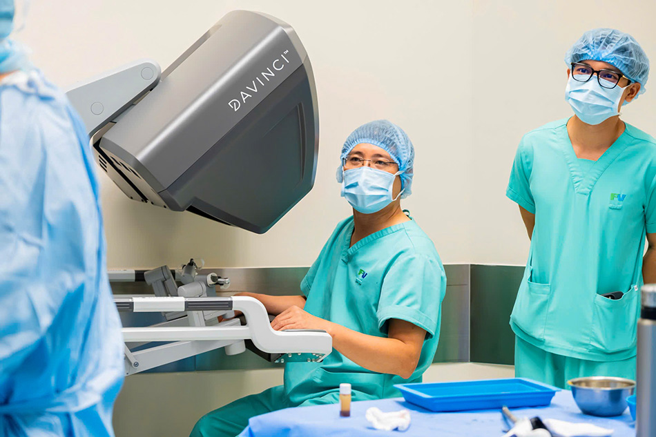
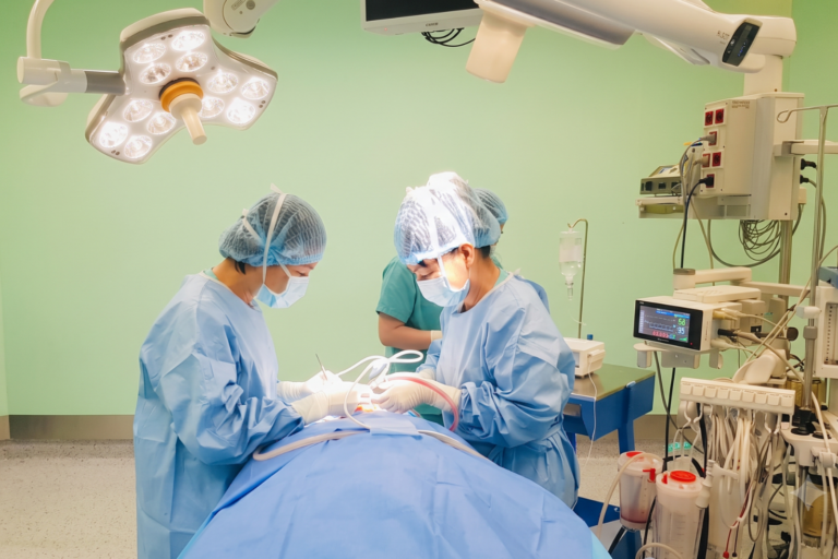
Dr Dang Dinh Minh Thanh, PhD, Specialist Level II and the surgical team performing lung tumour surgery on the patient. Photo: FV Hospital
Small lung nodule in a severely damaged lung
Mr Nguyen Van Hai, who lives in Ho Chi Minh City, had smoked approximately one packet of cigarettes a day for 40 years. He had contracted pulmonary tuberculosis around 30 years ago and also lives with diabetes.
Clinical evaluations and CT scans at FV Hospital revealed a suspected malignant lung nodule within heavily scarred and adherent lung tissue.
According to Dr Dang Dinh Minh Thanh, PhD, Specialist Level II, this was a clinically complex case. The patient had tuberculosis 30 years ago, which caused extensive adhesions throughout the lung, making it difficult to distinguish the boundaries between the lobes. The pleura was also fused to the chest wall. The surgery required not only removing the tumour, but also releasing these dense adhesions between the lung and the chest wall. We also had to minimise the risk of tearing the lung parenchyma, which was already fragile after years of damage.
Following clinical consultation, Mr Hai and his family agreed to proceed with da Vinci Xi robotic surgery under the care of Dr Dang Dinh Minh Thanh.
Nearly two hours of adhesiolysis before reaching the tumour
The first stage of the procedure involved freeing the lung from the chest wall. Due to the severe adhesions, the surgical team had to work meticulously, separating the layers of tissue between the lung and chest wall with great precision.
This step carried significant risks. The surface of the patient’s lung had multiple bullae (air-filled blisters). Any excessive force could rupture these blisters, leading to a pneumothorax (collapsed lung).
“The most demanding part of the adhesiolysis was around the lung apex and medial surface, adjacent to the mediastinum and major blood vessels. Even a minor error could put the patient’s life at risk,” explained Dr Thanh.
The da Vinci Xi surgical system allowed the surgeon to perform delicate dissections within a narrow anatomical space with high precision, while minimising unintended damage to the lung tissue. Nevertheless, due to the density of the adhesions, the adhesiolysis phase alone took nearly two hours.
Real-time identification of lung lobe boundaries using ICG technology
After freeing the lung from the chest wall, the team needed to determine the exact boundaries between the lung lobes. This presented another challenge because the tuberculosis sequelae had altered the lung’s natural anatomy.
To assist the procedure, Dr Thanh activated the ICG fluorescence imaging mode on the da Vinci Xi system. With this technology, the relevant lung tissue fluoresced, helping the surgeon clearly visualise anatomical boundaries in real time.
Combined with selective single-lung ventilation during anaesthesia, the team mapped the lobe margins and pinpointed the lesion. The da Vinci Xi system also helped the surgeon operate steadily in confined spaces by filtering out natural hand tremors. This allowed the team to access and excise the tumour while preserving healthy surrounding lung tissue.
Once the margins were clearly identified, the team excised the tumour. Pathological examination confirmed that the 17 mm lesion was lung cancer. As there were no signs of enlarged lymph nodes, the patient was diagnosed with stage IA lung cancer.
Under the treatment guidelines followed by the team, adjuvant therapy was not indicated following radical curative resection for this early-stage lung cancer.
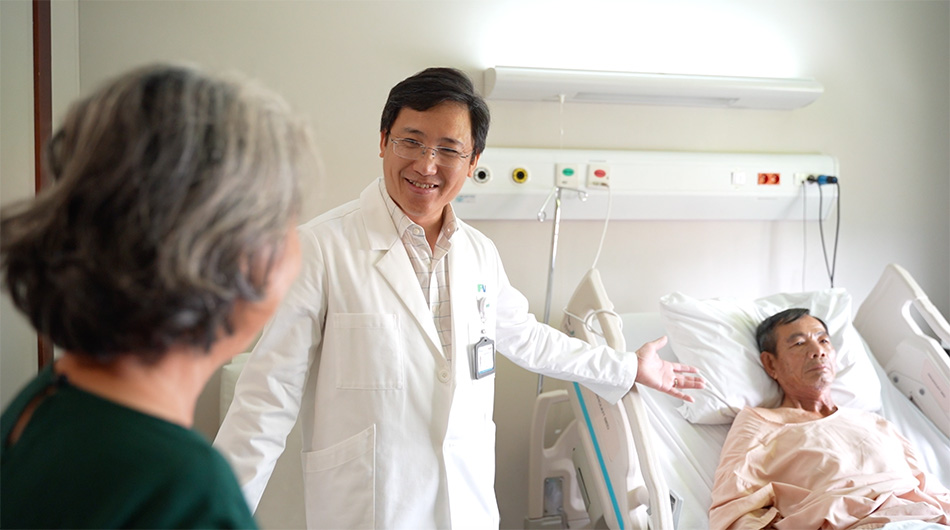
The patient recovered faster than expected after da Vinci Xi robotic surgery for lung cancer. Photo: FV Hospital
After surgery, Mr Hai’s respiratory function almost fully recovered within 24 hours. His stable condition provided great reassurance to him and his family.
“Throughout my treatment at FV, from customer service and consultation to paperwork, I have been thoroughly satisfied. The team is truly professional and dedicated,” shared Mr Hai.
Mr Hai’s case shows that tumour size is not the sole factor determining the complexity of surgery. For patients with lung damage caused by smoking or previous illnesses, a comprehensive assessment of lung condition and the selection of an appropriate surgical technique can play a crucial role in achieving a successful outcome.
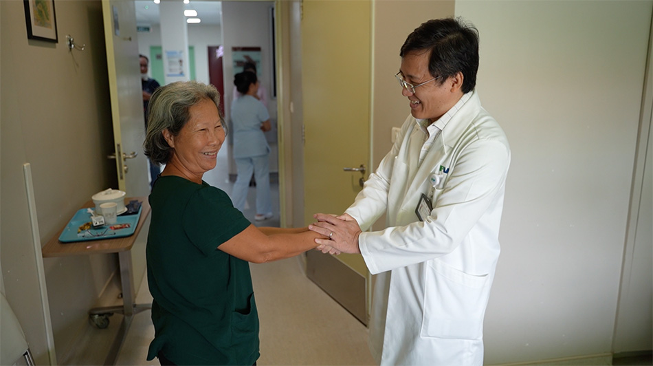
The patient’s relatives express their gratitude to Dr Thanh and the FV Hospital surgical team following the successful procedure. Photo: FV Hospital
For more information about da Vinci Xi robotic surgery, please contact FV Hospital at 06 Nguyen Luong Bang Street, Tan My Ward, Ho Chi Minh City (formerly District 7), or call (028) 3511 3333.
Related News
See moreRelated News
FV surgeons use robotic surgery to remove a 7cm tumour while preserving a female patient’s kidney
With the assistance of the da Vinci Xi robotic system, the FV Hospital surgical team successfully removed the entire 7cm tumour from the upper pole of the patient’s right kidney without removing the kidney. Large kidney tumour detected during a routine health check Nguyen Thi Lien, 56, from Quang Ngai, discovered a tumour in her […]

FV Surgeons Use Robotic Surgery to Remove Rectal Tumour, Ending an Elderly Man’s Nightly Suffering
For six months, a 62-year-old man from Lam Dong Province lived in distress, waking dozens of times each night to use the toilet. His fear of surgery led him to delay treatment, which allowed his rectal tumour to grow, nearly obstructing his bowel and invading his bladder. Delaying treatment led to a complex, advanced rectal […]

Minimally invasive surgery ends decades of sweaty palms for one patient
For over twenty years, 22-year-old Ta Duy Khang struggled with the embarrassment of cold, sweaty palms. The condition was so severe that he often avoided shaking hands. After a quick five-minute endoscopic thoracic sympathectomy at FV Hospital, his palms became dry and his confidence returned. Doctors at FV Hospital say that endoscopic thoracic surgery for […]
How 10 dental implants changed one patient’s life at FV Hospital
After years of tooth loss led to severe jawbone resorption and difficulty eating, specialists at FV Hospital restored the patient’s smile by placing 10 dental implants in one procedure. How years of tooth loss lead to jawbone loss For many years, Mr Lam Quang Kieu Phong (55, Ho Chi Minh City) experienced tooth loss. Some […]
FV surgeons use robotic surgery to remove a 7cm tumour while preserving a female patient’s kidney
FV Surgeons Use Robotic Surgery to Remove Rectal Tumour, Ending an Elderly Man’s Nightly Suffering
Minimally invasive surgery ends decades of sweaty palms for one patient

