



After a sudden toothache led to the discovery of a large mandibular cyst, a Ukrainian man spent six months searching for answers. His journey finally brought him to FV Hospital, where doctors crafted a two-stage treatment plan to preserve both his jawbone and teeth
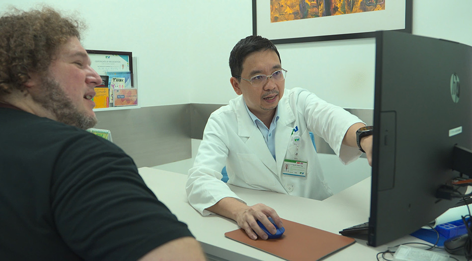
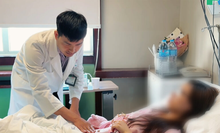
Dr Nguyen Thanh Tung, PhD, advising the patient on the treatment approach.
A six-month search for the right treatment
Mr Bohdan Vasiluk, a 37-year-old Ukrainian living in Da Nang, visited a dentist due to a toothache. A dental X-ray later revealed a large cystic lesion in his lower jawbone.
As this was beyond routine dental care, he was referred to a hospital with specialised expertise. Over the next six months, Bohdan visited multiple clinics and hospitals in Vietnam and traveled to Thailand, but could not find a suitable treatment plan.
A Vietnamese doctor then referred him to FV Hospital. Bohdan reached out to the hospital by email, sharing his medical history and scans. His case landed on the desk of Dr Nguyen Thanh Tung, PhD, Head of Dental & Maxillofacial Surgery at FV Hospital. After careful review, Dr Tung assured Bohdan that his condition could be treated. At last, after months of uncertainty, Bohdan saw a way forward.
A large cyst, yet almost no symptoms
On the surface, Bohdan’s face and mouth looked perfectly normal. He felt no numbness, no loss of sensation, and showed no signs of facial deformity.
However, 3D imaging revealed a lesion measuring approximately 56 mm in diameter that had destroyed a substantial portion of the lower jawbone, extending into the ramus of the mandible, whilst also displacing the inferior alveolar nerve canal and causing resorption of bone surrounding the roots of the lower left molar teeth.
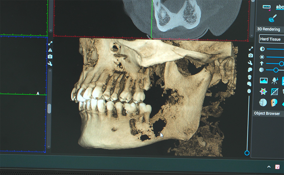
Imaging shows the approximately 56 mm mandibular cyst, causing severe bone destruction.
Dr Nguyen Thanh Tung, PhD, said cystic lesions of the jaw can progress silently. Some cases cause no obvious pain or swelling and are only discovered incidentally during a routine dental X-ray.
Medical reports have shown that jaw cysts can grow quite large before causing clear symptoms. As the lesion gets bigger, doctors must consider the risk of harming teeth, nerves, and bone, and in severe cases, the chance of the jaw breaking.
For Bohdan, the bone left in his jaw was very thin. Taking out the whole cyst in one surgery would have made it much more likely to damage his jawbone and teeth. But letting the cyst keep growing would make the bone even thinner.
Dr Tung had to find a way to treat the cyst while keeping as much bone and as many healthy teeth as possible.
Opting for a safer, two-stage treatment approach
After careful evaluation, Dr Nguyen Thanh Tung, PhD, MD decided on a conservative, two-stage treatment.
First, he performed cyst decompression, which means making a surgical opening in the cyst to release pressure. This helps the cyst shrink over time and allows new bone to grow.
Decompression is a well-established method for treating large jaw cysts. Research suggests it can shrink the lesion and help new bone form, paving the way for a less invasive surgery later if needed.
Bohdan’s decompression surgery took about an hour, led by Dr Tung and his team. Most of the lesion was removed, and a decompression tube was placed. Where the cyst pressed close to healthy teeth and nerves, the team worked with precision to protect these vital areas.
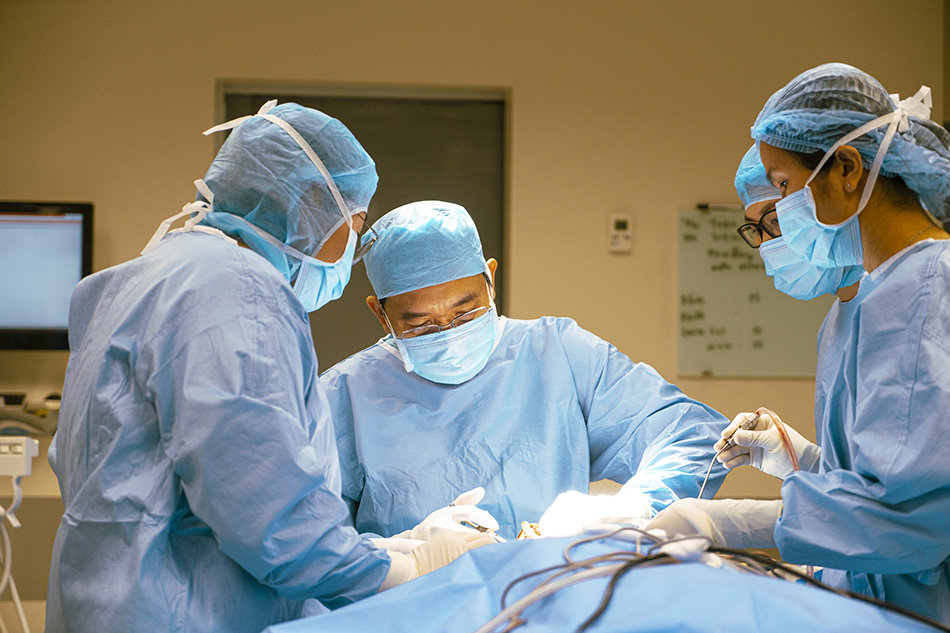
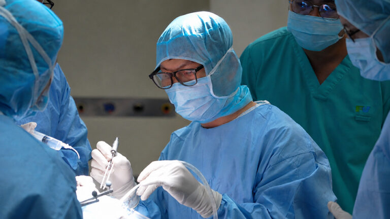
Dr Nguyen Thanh Tung, PhD, during the surgical procedure to treat the mandibular cyst in the Ukrainian patient.
After surgery, Bohdan learned how to care for the cavity with daily cleaning and returned for regular check-ups.
Just a month later, scans revealed promising news: the area was healing, and a delicate layer of new bone had started to grow. Should this regeneration continue favourably, Dr Tung anticipates that Bohdan could be assessed for the next stage of treatment in approximately one to two years, to address the remaining portion of the lesion.
Splitting treatment into two parts is a common strategy for large jaw cysts. Decompression often comes first, followed by complete removal of the cyst lining once the lesion has shrunk and bone has strengthened. The timing depends on the cyst’s type, size, and how well the bone heals, as seen on follow-up scans.
For Bohdan, what mattered most after surgery was not how far his wound had healed, but the fact that his own teeth had been preserved.
“Dr Tung is highly skilled, and everything went exactly according to plan. What mattered most to me was that he found a way to save my teeth. At every follow-up appointment, he told me the healing was progressing well, and that made me truly happy,” Bohdan said.
The importance of early detection for jaw cysts
According to Dr Nguyen Thanh Tung, Bohdan’s case shows that some jaw lesions can progress for long periods without symptoms. Anyone with impacted, malpositioned, or otherwise abnormal teeth or jaw should consult an oral and maxillofacial specialist and undergo imaging if indicated. X-rays can detect lesions deep within the bone that are not visible during examination.
For large lesions, surgeons must consider size, location, and impact on bone, teeth, and surrounding structures to determine the best treatment. In some cases, a staged approach can reduce surgical invasiveness and better preserve key anatomical structures.
At FV Hospital, the Oral and Maxillofacial Surgery Department provides diagnosis and treatment for the full range of oral and maxillofacial conditions, including complex lesions requiring a combination of clinical examination and diagnostic imaging to develop an appropriate treatment plan. For more information on specialist oral and maxillofacial treatment, please contact the Oral and Maxillofacial Surgery Department at FV Hospital, 6 Nguyen Luong Bang Street, Tan My Ward (formerly District 7), Ho Chi Minh City, or call (028) 3511 3333.
Related News
See moreRelated News
Abdominal pain misdiagnosed as digestive issue: 23-year-old woman diagnosed with ovarian torsion
While studying abroad in South Korea, N.T.T.M. (23) experienced unusual pain in her lower right abdomen. She sought treatment at a local hospital and received medication for a suspected digestive issue. After returning to Vietnam to visit her family, the pain recurred and worsened. On her friends’ advice, M. visited FV Hospital for evaluation. She […]
Advanced robotic surgery at FV Hospital removes tumour near aorta
Surgeons at FV Hospital used the da Vinci Xi robotic surgical system to successfully remove a fibrous tumour located in a difficult-to-access area next to the aorta in a 46-year-old woman. Ms Nguyen Thuy Phuong Dung recovered well after surgery to remove the fibrous tumour from her left chest. Image: FV When a subtle chest […]
Boy with ‘boneless arm’ and decade-long journey to find hope
A jump at the age of six left young D.V.C. with devastating consequences: a left forearm almost entirely devoid of skeletal architecture, facing the prospect of limb amputation through joint disarticulation. For more than ten years, hope faded as every attempt at recovery stalled. It was only through the intervention of the Children of Vietnam […]
Travelling halfway across the world for specialist spinal stenosis treatment
A Canadian woman who had nearly lost the ability to walk due to spinal canal stenosis was successfully treated at FV Hospital and was able to return home just one week after surgery. More than thirty years ago, Ms M.C. (49, Canada) underwent scoliosis correction surgery. In the past year, she developed severe back pain […]
Abdominal pain misdiagnosed as digestive issue: 23-year-old woman diagnosed with ovarian torsion
Advanced robotic surgery at FV Hospital removes tumour near aorta
Boy with ‘boneless arm’ and decade-long journey to find hope

